

Elif AtayT., Gülnur UzunE.
Exosomes classified as a subtype of extracellular vesicle (EV) and formed by endosomal way1-3, ~ have 30-150 nanometers (nm) diameter3-10 and spherical shape based on their scanning electron microscope (SEM)4,11-14; in addition, their intracellular biogenesis’ are unique, for this reason, they differ from other subtypes of EV15.
In 1981, even though Trams et al.4,16,17 defined exosomes as vesicles formed as a result of plasma membranes’ exfoliation, the most certain state of the “exosome” term was derived in 198718. Exosomes were first identified in the period of transferrin (Tfr) receptors’, which occurs as a result of reticulocytes’ maturation and elimination. Reticulocytes are subjected to strong cellular reprogramming in the first step of their maturation; towards the end of this process, Tfr receptors are released to the extracellular matrix with the help of multivesicular bodies (MVB), which have small vesicles11,19. Exosomes contain specific and non-specific proteins. Among tetraspanins20, one of the non-specific proteins, CD63 and CD81 antigens are the most abundant proteins on the ILV (intraluminal vesicles) membranes21. Tetraspanins keep in touch with different protein partners such as integrins and MHC (major histocompatibility complex) molecules, which is why they help large molecular complexes to unite20.
Exosomes secreted by various types of cells20,22,23; promise to be used as a tool for the future that allows early diagnosis of diseases 1,3,18,25 thanks to the advantage of their presence in biological fluids such as plasma, urine, seminal fluid, saliva, bronchial fluid, cerebrospinal fluid, breast milk, serum, amniotic fluid, synovial fluid, tear, lymph, bile, and gastric acid1,4,6,7,15,24.
In this review, aims to show the structural components of exosomes, and their biogenesis and give information about their therapeutic potential considering their importance in diagnosis as a biomarker, and the latest developments.
Exosome Structure and Its Molecular Interactions
Although exosomes are produced by the same cell, they are heterogeneous in terms of size and composition of components. Heat shock proteins (HSP), MVB biogenesis proteins (ALG-2 interacting protein (ALIX), tumor susceptibility gene 101 (TSG101)) and tetraspanins found in exosomes usually loaded on them and are used as exosome biomarkers7,26,27. Even though the biological function of fragmented nucleic acids (DNA, mRNA, miRNA, piRNA, snoRNA, snRNA, rRNA, tRNA, Y-RNA, scRNA) is not understood, it is known that they can be loaded onto exosomes too. Exosomes also contain lipids such as cholesterol, phosphatidylserine (PS), sphingomyelin, phosphatidic acid, sphingolipid, and ceramide; cholesterol, PS and sphingomyelin generally exist on exosome membrane26,27 (Figure 1)27.
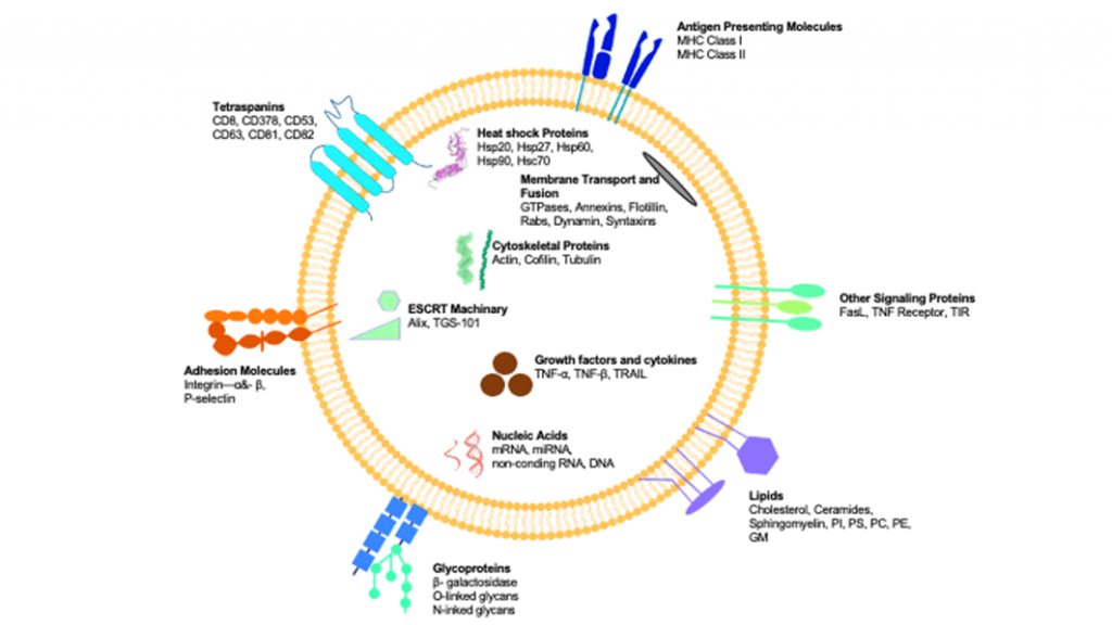
Figure 1. Structural components of an exosome27. Exosomes are formed by various proteins. Proteins such as transmembrane proteins like tetraspanins, antigen-presenting molecules, glycoproteins, and adhesion molecules; Hsc, cytoskeletal proteins, ESCRT (endosomal complexes for required transport) components, membrane transport, fusion proteins, growth factors, and cytokines are examined in the exosomes’ lumen. Hsp, heat shock proteins; TSG, tumor suppressor gene; TNF, tumor necrosis factor; TGF, transforming growth factor; TRAIL, a ligand that induces TNF-dependent apoptosis; FasL, Fas ligand.
In the process, exosomal vesicles occur via the budding of early endosomes’ restrictive membrane, which is converted into MVBs that are 100-1000 nm in size10,28,29, to the inwards7,15,24,30. Early endosomes and MVBs are responsible for cell materials’ traffic and endocytic functions7,11,13. At the end of the process, MVBs head towards the lysosome for degradation or merge with the plasma membrane of the cell in order to release the content to the extracellular matrix. MVBs have significant roles related to protein classification, recycling, storing, carrying, and releasing. Endosomal separating complex, which is necessary for ESCRT (endosomal complexes for required transport) mechanism, regulates MVB and exosome formation and release; although this mechanism is still not completely understood, it is known that growth factors can be a stimulant in MVBs’ formation and cell might arrange exosome formation according to its needs properly7.
According to the latest research about exosomes, they have roles in plenty of processes such as intercellular signaling15, coagulation, inflammation, and providing intracellular homeostasis. These vesicles, which have the ability to transfer messenger RNA (mRNA), microRNA (miRNA), protein, enzyme, and lipids6,7, are easily available via liquid biopsy1,6,15,24. Exosome-based liquid biopsy points out1,15 that exosomes have potential advantages, and they are promising biomarkers in the identifying level of diagnosis and prognosis of cancer and autoimmune, infectious, and neurodegenerative diseases6,11,24; moreover, stability and expression level that exosomes have is another factor that provides exosomes to be considered as ideal biomarkers31.
Exosomes have the advantage of lowering the possibility of being imprisoned in the microvascular system than main cells and it is thought that they do not have a malignancy risk, since they cannot divide; also, it is stated that exosomes are more durable since both they are more stable than in vivo cells, and they can be lyophilized without any lost activity and stored5,32,33.
Exosome Biogenesis
As exosome biogenesis might be beneficial for molecular biology in understanding mechanisms, intercellular signaling, and release into the extracellular matrix, it is also important for molecular and medical applications where these processes might be targeted34. Generally, biofunctions of exosomes are related to their biogenesis, and cargos vary depending on cellular origin and extracellular environment7.
In exosome biogenesis, which begins in the endosomal system, early endosomes are converted to late endosomes or MVBs by maturation; meanwhile, the endosomal membrane undergoes invagination10,14,21,29,31,35-38 (formation of a knuckle from the surface towards the inside)39 to form ILVs on the lumen of organelles.The membrane of MVBs buds towards the inner lumen in order to create lots of ILV. At the end, MVBs fuse with lysosome or cell membrane for dissociation, and ILVs are released to the extracellular matrix by exocytosis37,40-45,47. Each step of exosome biogenesis is mediated by multiple mechanisms that are highly variable depending on the cargo, cell type, and microenvironment and lead to heterogeneity of exosomes47 (Figure 2)46.
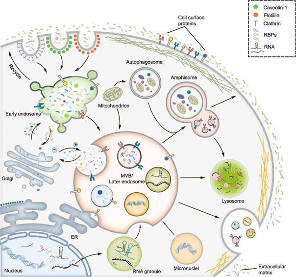
Figure 2. Exosome biogenesis mechanism46. MVBs are in the center of the exosome biogenesis process; it is generally produced by endocytosis, where different mechanisms mediate the processes of inward budding of the plasma membrane and formation of early endosomes. MVBs can interact with different organelles such as golgi, endoplasmic reticulum (ER), mitochondria, phagosome, and RNA; after completion of MVBs maturation, they can fuse with lysosomes to be digested or plasma membrane to free ILVs; in addition, MVBs can also merge with autophagosomes to form amphisomes.
EV traffic is controlled by Rab GTPases such as RAB14, RAB22, RAB27, and RAB37; besides, ILV formation has a role in MVBs and cell membranes’ fusion, and vesicles’ transportation processes17,35,42.
Biogenesis of MVBs is coordinated by ESCRT30,34. ESCRT mechanism has the function of exosomes’ formation and cargo hierarchy regulation29,38,40. The structure that consists of multi-protein complex named ESCRT-0, ESCRT-I, ESCRT-II, and ESCRT-III17,21,26,35 is generally classified under the cytosolic portion of the endosomal membrane in the process of classifying the selected protein based on ILVs34, and this complex structure can identify the cargos that can be found everywhere, vacuolar protein sorting 4 (VPS4), ALIX, and TSG10121,40. ESCRT proteins are also necessary for the point of targeting membrane proteins for lysosomal dissociation34.
ESCRT-0 participates in the assembly of proteins, which can be found everywhere, for clustering of cargo content and internalization. ESCRT-I/II undertakes the role of encouraging inward budding processes and cargo proteins’ enzymatic deubiquitination. ESCRT-III provides vesicle division and separation; adenosine triphosphatase (AAA + ATPase) is broken into pieces by VPS440. TSG101 found in the ESCRT-I complex causes the formation of a complex that binds to cargo proteins, and ESCRT-III complex oligomerization and formation by providing activation of the ESCRT-II complex. ESCRT-III is dissociated by an ATPase34. The most comprehensive research about ESCRTs in exosome biogenesis is carried out with RNAi (RNA interference) scanning which targets ESCRT and ESCRT related to proteins in HeLa cells35.
Studies show that exosome biogenesis continues in the existence of ESCRT inhibitors; this leads to consideration of the existence of ESCRT-independent pathways36,40. Both ESCRT-dependent and ESCRT-independent pathways play roles in exosome formation and controlling cargo arranging, and the amount of exosome secretion differs due to the selective exportation of original cells14,17,26,37,40,41. Protein uptake and cargo clustering are provided by both ESCRT-dependent and ESCRT-independent mechanisms14,48. In the ESCRT-dependent mechanism, the ESCRT-0 complex is responsible for protein collecting via ubiquitin or clathrin; controlling mechanisms of ESCRT I/II/III complexes. At the end of the process, ESCRT complexes are recycled for the next exosome biogenesis loop by breaking into pieces14,26,49. The ESCRT-independent mechanism contains syntenin, ALIX, and ESCRT-III. Syntenin collects proteins to form cargo clusters; then, ALIX and ESCRT-III arrange the budding process, and formation of ILV and exosome14,26,48,49.
Exosomes’ Therapeutic Potential and Alternative Areas of Use
Exosomes have the potential for angiogenesis and cancer metastasis, which are the two main aims of cancer therapy, thanks to their characteristic features such as molecular composition and critical immunogenicity45,50; besides, there is a great interest in exosomes for cancer treatment, since they can drain bioactive charge to cancer cells51. Exosomes that are double-layered, nano-sized, cell-free, and derived from hosts, have the potential of transporting charges to targeted cells, which are determined via the high level of specific biodistribution and low immunogenicity52. In disease diagnosis and treatment, exosome-based strategies are evaluated as a powerful tool for the depletion of tumor-derived exosomes by using exosome inhibitors, application of exosomes derived from specific cell types for therapeutic purposes, and delivery of antineoplastic agents to selective target sites53.
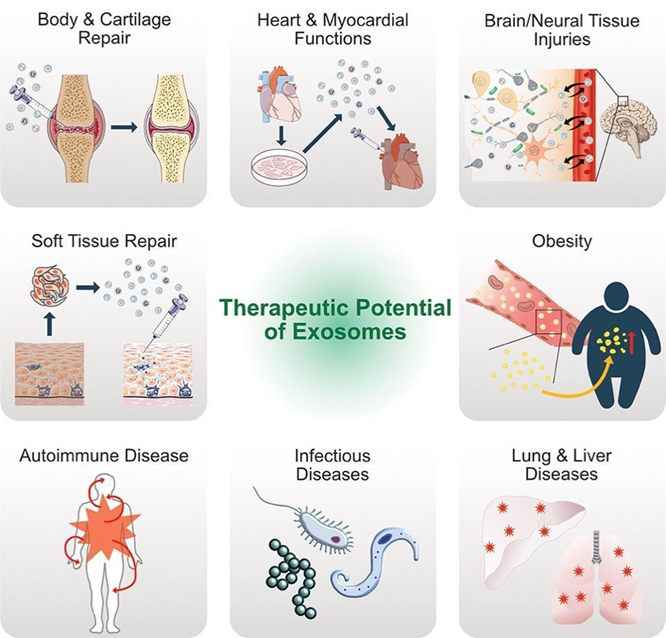
Figure 3. Therapeutic Potential of Exosomes45. Exosomes associated with many processes including central nervous system diseases (CNS), myocardial ischemia/circulatory damage, immune responses, viral pathogenicity, cardiovascular diseases, and cancer progression1,18,45 (Figure 3) are quite easily available nowadays and have the advantage of being measurable biomarker; in addition to this, it has great importance for gene delivery vehicles and regenerative medicine because of its ability to cross blood-brain barrier34. The identified therapeutic advantages of exosomes have significantly increased the interest in exosome-based treatment modalities examined in experimental studies for different pathologies such as sepsis, diabetes, cancer, wound healing, and stroke18.
From the beginning of carcinogenesis, the complex exosomal communication network built between tumor cells and healthy intercellular plays an active role in every step of cancer. Tumor cells stimulate a suitable microenvironment that supports tumor growth via mature cell proliferation and running away from apoptosis mechanisms, and exosome-mediated mechanisms are utilized for this54,55. In cancer, exosomes provide an environment for new vessel formations and contribute these cells to being metabolically programmed again by providing nutrition and oxygen for the continuous proliferation of neoplastic cells55-57. Additionally, cancer exosome subsets critically contribute to cancer development and progression; thus, understanding mechanisms in their biogenesis provides new options and research areas for therapeutic strategies. Exosome-based modalities developed for cancer therapy are examined in four categories51-58 as use of exosomes that are naturally derived from immune cells to suppress cancer cells43, inhibition of cancer-originated exosomes release, use of exosomes as gene carriers58, and use of exosomes as drug carriers.
Stated requirements for the therapeutic effects of exosomes that enable the development of alternative strategies as drugs and gene delivery vehicles are lined up as encapsulating the required amount of drug, having long-term intrinsic stability in size, structure, and bioactivity of the therapeutic agent during circulation, being capable of escaping from macrophages, being non-toxic or having low toxicity, being biocompatible with the immune response, and being non-immunogenic31.
Future Perspectives and Difficulties in Exosome Therapy
Exosome biogenesis and related mechanisms have some restrictions. The mechanisms that underlie MVBs’ differentiation towards dissociation or fusion with cell membranes are not known and need to be studied. The mechanism that the endocytic system arranges MVBs’ percentage towards cell membrane and fusion is another restriction. In addition, from ILVs to MVBs, how the cargo content classification is regulated is still not fully illuminated21.
Even though there are a lot of successful experimental studies that are about the therapeutic effects of exosomes, before being able to use exosome-based therapeutics, many difficulties need to be solved primarily isolation-purification, cell source, large-scaled production, dosage-potential, uptake-distribution, and safety, due to the much more need of clinical studies60. Although there are different strategies for exosome isolation, which have great importance, the gold standard is evaluated as centrifuge-based methods; in addition, ultrafiltration is one of the most important modalities for size-based isolation and is usually used with ultracentrifugation. Microfluidic-based technologies have recently become important techniques that benefit from both the physical and biochemical features of exosomes. Since different exosome isolation modalities can affect the analysis, it is specifically important to choose suitable isolation methods carefully61.
Traditional cell culture methods that are preferred in exosome formation have disadvantages in terms of cost and time. Even though 2-dimensional (2D) culture techniques are generally used in the process of obtaining exosomes, they produce exosomes at low levels. On the other hand, the use of 3-dimensional (3D) culturing techniques or bioreactors increases efficiency in productivity, but these approaches also require more environment and more frequent transitions62,63. In the case of larger-scaled methods, heterogeneity in the cells and depending on this in the exosomes is still a problem although cell culture surface area is maximized for expansion. Determination of exosomes’ dosage and strength for clinical use is also evaluated among difficulties because exosomes are heterogeneous in different progenitor cells and animal models used for exosome treatment; moreover, there are differences in cellular features and evaluating62,64,65 The large-scale of exosomes’ production, expansion, and distribution are also faced as important difficulties in regenerative medicine. As purification and development in production continue, it is expected exosomes will be more target-specific and tissue-specific in regenerative medicine66. Since exosomes taken by different types of cells represent different features, when tissue is selected and methods are used for delivering exosomes to the targeted tissue, standards can be optimized in a more efficient way. Although therapeutic characteristics of exosomes that are produced by mesenchymal stem cells (MSCs), the American Food and Drug Administration (FDA) does not approve converting into clinical trials due to the heterogeneity in exosomes, the complexity of validation techniques besides cell sources and isolation; this emphasizes the importance of conducting comprehensive research about exosome stability, safety, efficacy, and quality in bioliquids62. When continuous development of exosome-based delivery systems, advancement, and optimization of gene editing tools are handled together, targeted gene therapies are emphasized and they have the potential of accessible treatments for complex diseases in the future. Today, even though there is a significant development in the large-scale production of exosomes or drug-loaded exosomes66, there are still difficulties in the practical use of exosomes17.
Considering its therapeutic potential for diseases, use in drug delivery systems, and advances in regenerative medicine, exosomes, which have a great development area, are thought to bring along innovative approaches and clinical validation studies against current difficulties and limitations as their heterogeneous structure continues to be solved. Having great potential in the biomedical field and being considered as new-generation nanomaterials provide a continuous increase in research.
References:
1.Kalluri, R., & LeBleu, V. S. (2020). The biology, function, and biomedical applications of exosomes. Science, 367(6478). https://doi.org/10.1126/science.aau6977
2. Blanc, L., De Gassart, A., Géminard, C., Bette-Bobillo, P., & Vidal, M. (2005). Exosome release by reticulocytes – An integral part of the red blood cell differentiation system. Blood Cells, Molecules, and Diseases, 35(1), 21–26. https://doi.org/10.1016/j.bcmd.2005.04.008
3.Jan, A. T., Rahman, S., Badierah, R., Lee, E. J., Mattar, E. H., Redwan, E. M., & Choi, I. (2021). Expedition into exosome biology: A perspective of progress from discovery to therapeutic development. Cancers, 13(5), 1–25. https://doi.org/10.3390/cancers13051157
4.Li, Y., Meng, L., Li, B., Li, Y., Shen, T., & Zhao, B. (2022). The Exosome Journey: From Biogenesis to Regulation and Function in Cancers. Journal of Oncology, 2022. https://doi.org/10.1155/2022/9356807
5.Zipkin, M. (2019). Exosome redux. Nature Biotechnology, 37(12), 1395–1400. https://doi.org/10.1038/s41587-019-0326-5
6.Boriachek, K., Islam, M. N., Möller, A., Salomon, C., Nguyen, N. T., Hossain, M. S. A., Yamauchi, Y., & Shiddiky, M. J. A. (2018). Biological Functions and Current Advances in Isolation and Detection Strategies for Exosome Nanovesicles. Small, 14(6), 1–21. https://doi.org/10.1002/smll.201702153
7.Doyle, L. M., & Wang, M. Z. (2019). Overview of extracellular vesicles, their origin, composition, purpose, and methods for exosome isolation and analysis. Cells, 8(7), 29–39. https://doi.org/10.3390/cells8070727
8.Sun, W., Luo, J. D., Jiang, H., & Duan, D. D. (2018). Tumor exosomes: A double-edged sword in cancer therapy. Acta Pharmacologica Sinica, 39(4), 534–541. https://doi.org/10.1038/aps.2018.17
9.Lee, Y., Morrison, B. M., Li, Y., Lengacher, S., Farah, M. H., Hoffman, P. N., Liu, Y., Tsingalia, A., Jin, L., Zhang, P. W., Pellerin, L., Magistretti, P. J., & Rothstein, J. D. (2012). Oligodendroglia metabolically support axons and contribute to neurodegeneration. Nature, 487(7408), 443–448. https://doi.org/10.1038/nature11314
10.Rashed, M. H., Bayraktar, E., Helal, G. K., Abd-Ellah, M. F., Amero, P., Chavez-Reyes, A., & Rodriguez-Aguayo, C. (2017). Exosomes: From garbage bins to promising therapeutic targets. International Journal of Molecular Sciences, 18(3). https://doi.org/10.3390/ijms18030538
11.Rajagopal, C., & Harikumar, K. B. (2018). The origin and functions of exosomes in cancer. Frontiers in Oncology, 8(MAR). https://doi.org/10.3389/fonc.2018.00066
12.Raposo, G., & Stoorvogel, W. (2013). Extracellular vesicles: Exosomes, microvesicles, and friends. Journal of Cell Biology, 200(4), 373–383. https://doi.org/10.1083/jcb.201211138
13. Von Bartheld, C. S., & Altick, A. L. (2011). Multivesicular bodies in neurons: Distribution, protein content, and trafficking functions. Progress in Neurobiology, 93(3), 313–340. https://doi.org/10.1016/j.pneurobio.2011.01.003
14.Jiang, P., Zhang, S., Cheng, C., Gao, S., Tang, M., Lu, L., Yang, G., & Chai, R. (2020). The Roles of Exosomes in Visual and Auditory Systems. Frontiers in Bioengineering and Biotechnology, 8(June), 1–11. https://doi.org/10.3389/fbioe.2020.00525
15.Liga, A., Vliegenthart, A. D. B., Oosthuyzen, W., Dear, J. W., & Kersaudy-Kerhoas, M. (2015). Exosome isolation: A microfluidic road-map. Lab on a Chip, 15(11), 2388–2394. https://doi.org/10.1039/c5lc00240k
16.Trams, E. G., Lauter, C. J., Norman Salem, J., & Heine, U. (1981). Exfoliation of membrane ecto-enzymes in the form of micro-vesicles. BBA – Biomembranes, 645(1), 63–70. https://doi.org/10.1016/0005-2736(81)90512-5
17. Liao, W., Du, Y., Zhang, C., Pan, F., Yao, Y., Zhang, T., & Peng, Q. (2019). Exosomes: The next generation of endogenous nanomaterials for advanced drug delivery and therapy. Acta Biomaterialia, 86, 1–14.https://doi.org/10.1016/j.actbio.2018.12.045
18.Sancho-Albero, M., Navascués, N., Mendoza, G., Sebastián, V., Arruebo, M., Martín-Duque, P., & Santamaría, J. (2019). Exosome origin determines cell targeting and the transfer of therapeutic nanoparticles towards target cells. Journal of Nanobiotechnology, 17(1), 1–13. https://doi.org/10.1186/s12951-018-0437-z
19.Harding, C., Heuser, J., & Stahl, P. (1983). Receptor-mediated endocytosis of transferrin and recycling of the transferrin receptor in rat reticulocytes. The Journal of Cell Biology, 97(2), 329–339. https://doi.org/10.1083/jcb.97.2.329
20. Deb, A., Gupta, S., & Mazumder, P. B. (2021). Exosomes: A new horizon in modern medicine. Life Sciences, 264(October 2020), 118623. https://doi.org/10.1016/j.lfs.2020.118623
21. Paskeh, M. D. A., Entezari, M., Mirzaei, S., Zabolian, A., Saleki, H., Naghdi, M. J., Sabet, S., Khoshbakht, M. A., Hashemi, M., Hushmandi, K., Sethi, G., Zarrabi, A., Kumar, A. P., Tan, S. C., Papadakis, M., Alexiou, A., Islam, M. A., Mostafavi, E., & Ashrafizadeh, M. (2022). Emerging role of exosomes in cancer progression and tumor microenvironment remodeling. In Journal of Hematology and Oncology (Vol. 15, Issue 1). BioMed Central. https://doi.org/10.1186/s13045-022-01305-4
22.Li, X., Corbett, A. L., Taatizadeh, E., Tasnim, N., Little, J. P., Garnis, C., Daugaard, M., Guns, E., Hoorfar, M., & Li, I. T. S. (2019). Challenges and opportunities in exosome research-Perspectives from biology, engineering, and cancer therapy. APL Bioengineering, 3(1). https://doi.org/10.1063/1.5087122
23.LeBleu, V. S., & Kalluri, R. (2020). Exosomes as a Multicomponent Biomarker Platform in Cancer. Trends in Cancer, 6(9), 767–774. https://doi.org/10.1016/j.trecan.2020.03.007
24. Hu, Q., Su, H., Li, J., Lyon, C., Tang, W., Wan, M., & Hu, T. Y. (2020). Clinical applications of exosome membrane proteins. Precision Clinical Medicine, 3(1), 54–66. https://doi.org/10.1093/pcmedi/pbaa007
25. Lin, J., Li, J., Huang, B., Liu, J., Chen, X., Chen, X. M., Xu, Y. M., Huang, L. F., & Wang, X. Z. (2015). Exosomes: Novel Biomarkers for Clinical Diagnosis. Scientific World Journal, 2015. https://doi.org/10.1155/2015/657086
26.Song, Y., Kim, Y., Ha, S., Sheller-Miller, S., Yoo, J., Choi, C., & Park, C. H. (2021). The emerging role of exosomes as novel therapeutics: Biology, technologies, clinical applications, and the next. American Journal of Reproductive Immunology, 85(2), 1–10. https://doi.org/10.1111/aji.13329
27. Gurung, S., Perocheau, D., Touramanidou, L., & Baruteau, J. (2021). The exosome journey: from biogenesis to uptake and intracellular signalling. Cell Communication and Signaling, 19(1), 1–19. https://doi.org/10.1186/s12964-021-00730-1
28. Gurunathan, S., Kang, M. H., Jeyaraj, M., Qasim, M., & Kim, J. H. (2019). Review of the isolation, characterization, biological function, and multifarious therapeutic approaches of exosomes. Cells, 8(4). https://doi.org/10.3390/cells8040307
29. Zhang, Y., Liu, Y., Liu, H., & Tang, W. H. (2019). Exosomes: Biogenesis, biologic function and clinical potential. Cell and Bioscience, 9(1), 1–18. https://doi.org/10.1186/s13578-019-0282-2
30. Bebelman, M. P., Smit, M. J., Pegtel, D. M., & Baglio, S. R. (2018). Biogenesis and function of extracellular vesicles in cancer. Pharmacology and Therapeutics, 188, 1–11. https://doi.org/10.1016/j.pharmthera.2018.02.013
31. Chung, I. M., Rajakumar, G., Venkidasamy, B., Subramanian, U., & Thiruvengadam, M. (2020). Exosomes: Current use and future applications. Clinica Chimica Acta, 500(October 2019), 226–232. https://doi.org/10.1016/j.cca.2019.10.022
32. Marbán E. (2018). The Secret Life of Exosomes: What Bees Can Teach Us About Next-Generation Therapeutics. Journal of the American College of Cardiology, 71(2), 193–200.https://doi.org/10.1016/j.jacc.2017.11.013
33. Akers, J. C., Ramakrishnan, V., Yang, I., Hua, W., Mao, Y., Carter, B. S., & Chen, C. C. (2016). Optimizing preservation of extracellular vesicular miRNAs derived from clinical cerebrospinal fluid. Cancer biomarkers : section A of Disease markers, 17(2), 125–132. https://doi.org/10.3233%2FCBM-160609
34.Urbanelli, L., Magini, A., Buratta, S., Brozzi, A., Sagini, K., Polchi, A., Tancini, B., & Emiliani, C. (2013). Signaling pathways in exosomes biogenesis, secretion and fate. Genes, 4(2), 152–170. https://doi.org/10.3390/genes4020152
35.Hessvik, N. P., & Llorente, A. (2018). Current knowledge on exosome biogenesis and release. Cellular and Molecular Life Sciences, 75(2), 193–208. https://doi.org/10.1007/s00018-017-2595-9
36.Anakor, E., Le Gall, L., Dumonceaux, J., Duddy, W. J., & Duguez, S. (2021). Exosomes in ageing and motor neurone disease: Biogenesis, uptake mechanisms, modifications in disease and uses in the development of biomarkers and therapeutics. Cells, 10(11). https://doi.org/10.3390/cells10112930
37. Abels, E. R., & Breakefield, X. O. (2016). Introduction to Extracellular Vesicles: Biogenesis, RNA Cargo Selection, Content, Release, and Uptake. Cellular and Molecular Neurobiology, 36(3), 301–312. https://doi.org/10.1007/s10571-016-0366-z
38. Jella, K. K., Nasti, T. H., Li, Z., Malla, S. R., Buchwald, Z. S., & Khan, M. K. (2018). Exosomes, their biogenesis and role in inter-cellular communication, tumor microenvironment and cancer immunotherapy. Vaccines, 6(4). https://doi.org/10.3390/vaccines6040069
39.https://doi.org/10.1016/B978-0-12-391062-2.00018-8
40.Jiang, L., Gu, Y., Du, Y., & Liu, J. (2019). Exosomes: Diagnostic Biomarkers and Therapeutic Delivery Vehicles for Cancer. Molecular Pharmaceutics, 16(8), 3333–3349. https://doi.org/10.1021/acs.molpharmaceut.9b00409
41. Jadli, A. S., Ballasy, N., Edalat, P., & Patel, V. B. (2020). Inside(sight) of tiny communicator: exosome biogenesis, secretion, and uptake. Molecular and Cellular Biochemistry, 467(1–2), 77–94. https://doi.org/10.1007/s11010-020-03703-z
42.Beshbishy, A. M., Alghamdi, S., Onyiche, T. G. E., Zahoor, M., Rivero-Perez, N., Zaragoza-Bastida, A., Ghorab, M. A., Meshaal, A. K., El-Esawi, M. A., Hetta, H. F., & El-Saber Batiha, G. (2020). Biogenesis, biologic function and clinical potential of exosomes in different diseases. Applied Sciences (Switzerland), 10(13). https://doi.org/10.3390/app10134428
43. Behera, J., & Tyagi, N. (2018). Exosomes: mediators of bone diseases, protection, and therapeutics potential. Oncoscience, 5(5–6), 181–195. https://doi.org/10.18632/oncoscience.421
44.Pan, B. T., & Johnstone, R. M. (1983). Fate of the transferrin receptor during maturation of sheep reticulocytes in vitro: Selective externalization of the receptor. Cell, 33(3), 967–978. https://doi.org/10.1016/0092-8674(83)90040-5
45.Gurunathan, S., Kang, M. H., & Kim, J. H. (2021). A comprehensive review on factors influences biogenesis, functions, therapeutic and clinical implications of Exosomes. International Journal of Nanomedicine, 16, 1281–1312. https://doi.org/10.2147/IJN.S291956
46. Han, Q. F., Li, W. J., Hu, K. S., Gao, J., Zhai, W. L., Yang, J. H., & Zhang, S. J. (2022). Exosome biogenesis: machinery, regulation, and therapeutic implications in cancer. Molecular Cancer, 21(1), 1–26. https://doi.org/10.1186/s12943-022-01671-0
47. Van Niel, G., D’Angelo, G., & Raposo, G. (2018). Shedding light on the cell biology of extracellular vesicles. Nature Reviews Molecular Cell Biology, 19(4), 213–228. https://doi.org/10.1038/nrm.2017.125
48. Colombo, M., Raposo, G., & Théry, C. (2014). Biogenesis, secretion, and intercellular interactions of exosomes and other extracellular vesicles. Annual Review of Cell and Developmental Biology, 30, 255–289. https://doi.org/10.1146/annurev-cellbio-101512-122326
49.Nam, G. H., Choi, Y., Kim, G. B., Kim, S., Kim, S. A., & Kim, I. S. (2020). Emerging Prospects of Exosomes for Cancer Treatment: From Conventional Therapy to Immunotherapy. Advanced Materials, 32(51). https://doi.org/10.1002/adma.202002440
50.Von Schulze, A., & Deng, F. (2020). A review on exosome-based cancer therapy. Journal of Cancer Metastasis and Treatment, 2020. https://doi.org/10.20517/2394-4722.2020.79
51.He, C., Zheng, S., Luo, Y., & Wang, B. (2018). Exosome theranostics: Biology and translational medicine. Theranostics, 8(1), 237–255. https://doi.org/10.7150/thno.21945
52. Wang, X., Tian, L., Lu, J., & Ng, I. O. L. (2022). Exosomes and cancer – Diagnostic and prognostic biomarkers and therapeutic vehicle. Oncogenesis, 11(1), 1–12. https://doi.org/10.1038/s41389-022-00431-5
53. Meehan, K., & Vella, L. J. (2016). The contribution of tumour-derived exosomes to the hallmarks of cancer. Critical Reviews in Clinical Laboratory Sciences, 53(2), 121–131. https://doi.org/10.3109/10408363.2015.1092496
54. Ruivo, C. F., Adem, B., Silva, M., & Melo, S. A. (2017). The biology of cancer exosomes: Insights and new perspectives. Cancer Research, 77(23), 6480–6488. https://doi.org/10.1158/0008-5472.CAN-17-0994
55. Fong, M. Y., Zhou, W., Liu, L., Alontaga, A. Y., Chandra, M., Ashby, J., Chow, A., O’Connor, S. T. F., Li, S., Chin, A. R., Somlo, G., Palomares, M., Li, Z., Tremblay, J. R., Tsuyada, A., Sun, G., Reid, M. A., Wu, X., Swiderski, P., … Wang, S. E. (2015). Breast-cancer-secreted miR-122 reprograms glucose metabolism in premetastatic niche to promote metastasis. Nature Cell Biology, 17(2), 183–194. https://doi.org/10.1038/ncb3094
56. Skog, J., Würdinger, T., van Rijn, S., Meijer, D. H., Gainche, L., Curry, W. T., Carter, B. S., Krichevsky, A. M., & Breakefield, X. O. (2008). Glioblastoma microvesicles transport RNA and proteins that promote tumour growth and provide diagnostic biomarkers. Nature Cell Biology, 10(12), 1470–1476. https://doi.org/10.1038/ncb1800
57. Lin, Q., Qu, M., Zhou, B., Patra, H. K., Sun, Z., Luo, Q., Yang, W., Wu, Y., Zhang, Y., Li, L., Deng, L., Wang, L., Gong, T., He, Q., Zhang, L., Sun, X., & Zhang, Z. (2019). Exosome-like nanoplatform modified with targeting ligand improves anti-cancer and anti-inflammation effects of imperialine. Journal of Controlled Release, 311–312(September), 104–116. https://doi.org/10.1016/j.jconrel.2019.08.037
58.Pullan, J. E., Confeld, M. I., Osborn, J. K., Kim, J., Sarkar, K., & Mallik, S. (2019). Exosomes as Drug Carriers for Cancer Therapy [Review-article]. Molecular Pharmaceutics, 16(5), 1789–1798. https://doi.org/10.1021/acs.molpharmaceut.9b00104
59. Kučuk, N., Primožič, M., Knez, Ž., & Leitgeb, M. (2021). Exosomes engineering and their roles as therapy delivery tools, therapeutic targets, and biomarkers. International Journal of Molecular Sciences, 22(17). https://doi.org/10.3390/ijms22179543
60. Wei, H., Chen, Q., Lin, L., Sha, C., Li, T., Liu, Y., Yin, X., Xu, Y., Chen, L., Gao, W., Li, Y., & Zhu, X. (2021). Regulation of exosome production and cargo sorting. International journal of biological sciences, 17(1), 163–177. https://doi.org/10.7150%2Fijbs.53671
61. Ahmadi, M., & Rezaie, J. (2021). Ageing and mesenchymal stem cells derived exosomes: Molecular insight and challenges. Cell Biochemistry and Function, 39(1), 60–66. https://doi.org/10.1002/cbf.3602
62. Phan, J., Kumar, P., Hao, D., Gao, K., Farmer, D., & Wang, A. (2018). Engineering mesenchymal stem cells to improve their exosome efficacy and yield for cell-free therapy. Journal of extracellular vesicles, 7(1), 1522236. https://doi.org/10.1080/20013078.2018.1522236
63. Kimiz-Gebologlu, I., & Oncel, S. S. (2022). Exosomes: Large-scale production, isolation, drug loading efficiency, and biodistribution and uptake. Journal of controlled release : official journal of the Controlled Release Society, 347, 533–543. https://doi.org/10.1016/j.jconrel.2022.05.027
64. Haraszti, R. A., Miller, R., Stoppato, M., Sere, Y. Y., Coles, A., Didiot, M. C., Wollacott, R., Sapp, E., Dubuke, M. L., Li, X., Shaffer, S. A., DiFiglia, M., Wang, Y., Aronin, N., & Khvorova, A. (2018). Exosomes Produced from 3D Cultures of MSCs by Tangential Flow Filtration Show Higher Yield and Improved Activity. Molecular therapy : the journal of the American Society of Gene Therapy, 26(12), 2838–2847. https://doi.org/10.1016/j.ymthe.2018.09.015
65. Popowski, K., Lutz, H., Hu, S., George, A., Dinh, P. U., & Cheng, K. (2020). Exosome therapeutics for lung regenerative medicine. Journal of Extracellular Vesicles, 9(1). https://doi.org/10.1080/20013078.2020.1785161 66. Duan, L., Xu, L., Xu, X., Qin, Z., Zhou, X., Xiao, Y., Liang, Y., & Xia, J. (2021). Exosome-mediated delivery of gene vectors for gene therapy. Nanoscale, 13(3), 1387–1397. https://doi.org/10.1039/d0nr07622h