
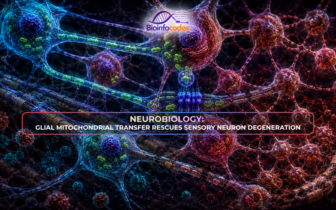
Primary sensory neurons located in the dorsal root ganglia (DRG) possess long peripheral axons that can extend up to one meter, creating high bioenergetic demands that require continuous mitochondrial function. Because mitochondrial dysfunction contributes to axonal degeneration and chronic pain, understanding how these neurons maintain their mitochondrial population is essential for neurobiology and peripheral nerve physiology.
Although sensory neurons rely heavily on mitochondrial energy production, the mechanisms supporting mitochondrial maintenance under physiological and pathological conditions remain unclear. Satellite glial cells (SGCs), which closely surround sensory neuron cell bodies within the DRG, are positioned to interact directly with neurons. This study investigated whether SGCs transfer mitochondria to DRG sensory neurons, identified the molecular and structural mechanisms involved, examined whether the process is activity-dependent, and evaluated its role in peripheral neuropathy.
The researchers used primary cell co-cultures, ex vivo DRG tissue, and transgenic mouse models. Fluorescently labelled SGC mitochondria were tracked during co-culture with sensory neurons, while pharmacological inhibitors were applied to determine transport mechanisms. Scanning and transmission electron microscopy (SEM and TEM) identified intercellular bridges, whereas MitoTag mice, single-nucleus RNA sequencing, calcium imaging, and mitochondrial or cell transplantation experiments were used to examine mitochondrial transfer in models of chemotherapy-induced and diabetic peripheral neuropathy.
The results showed that SGCs transfer mitochondria to DRG sensory neurons through tunneling nanotubes (TNTs), endocytosis, and gap junctions in vitro, ex vivo, and in vivo. SEM and TEM confirmed TNT-like structures containing mitochondria between SGCs and neurons. Myosin 10 (MYO10) was highly expressed in human SGCs, and its knockdown or deletion reduced TNT formation and mitochondrial transfer. Mechanistically, co-culturing with SGCs reduced paclitaxel-induced neuronal reactive oxygen species (ROS) and hyperexcitability, effects that were lost following CytoB treatment. In vivo, mitochondrial transfer occurred predominantly from SGCs to medium-to-large sensory neurons, increased after nerve injury, and decreased upon neuronal activity blockade or MYO10 deficiency. In addition, SGCs from human DRG donors with diabetes exhibited reduced MYO10 expression and impaired mitochondrial transfer. Transplantation of healthy human SGCs reduced mechanical hypersensitivity and improved mitochondrial function in mouse models of chemotherapy-induced and diabetic peripheral neuropathy. Direct transfer of mitochondria from healthy human SGCs also reduced neuropathic pain and increased intraepidermal nerve fiber density.
Overall, the study identifies a neuro-glial mechanism in which SGCs support neuronal mitochondrial function through MYO10-dependent TNTs. Impaired mitochondrial transfer contributes to nerve degeneration and pain hypersensitivity, while restoring this process improves peripheral nerve function, highlighting organelle transfer as a potential therapeutic strategy for peripheral neuropathies.
Author: Ezel Özerdem
Editor: Nehir Necem Ünlü
Reference:
Xu, J., Li, Y., Novak, C., Lee, M., Yan, Z., Bang, S., McGinnis, A., Chandra, S., Zhang, V., He, W., Lechler, T., Rodriguez Salazar, M. P., Eroglu, C., Becker, M. L., Velmeshev, D., Cheney, R. E., & Ji, R.-R. (2026). Mitochondrial transfer from glia to neurons protects against peripheral neuropathy. Nature. https://doi.org/10.1038/s41586-025-09896-x
-Bioinfocodes Scientific News Service-
News articles prepared by our team members, reviewing and compiling scientific research
published in journals with and impact factor greater than 20 (click here for the list)