

Ebru KavaklıT., Nida Dereli ÇalışkanE., Damla PehlivanR.
Introduction
Reoviruses taxonomically belong to the Reoviridae family and the Sedovirinae subfamily. Their names are derived from the initials of Respiratory Enteric Orphan (REO) because they represent respiratory and enteric viruses1. Reoviruses have an icosahedral capsid and a double-stranded Ribonucleic Acid (dsRNA) genome with 9-12 segments, approximately 16–27 kilobase pair (kbp), and 60–80 nanometer (nm) wide1,2. Unlike Adenoviruses and Herpes simplex viruses, Reoviruses have dsRNA instead of dsDNA. The RNA backbone of the genome reduces the genomic stability and makes the genome prone to mutations, complicating genetic applications3.
X-ray crystallography, 3D cryogenic electron microscopy (Cryo-EM) of the particles, and electron micrograph approaches are used to study the morphologies of members of the Reoviridae family (Figure 1)1. Although Reoviridae family members have similar morphologies in general, these approaches can be used to detect minor differences between them. Reoviruses can infect various organisms, such as mammals, birds, reptiles, amphibians, fish, mollusks, crustaceans, plants, fungi, and protists4.
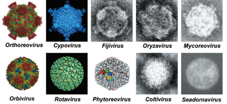
Figure 1. The comparison of two different core particle morphologies seen in the members of Spinareovirinae and Sedoreovirinae subfamilies.
Reoviruses have 15 genera and are divided into two subfamilies: Spinareovirinae and Sedoreovirinae5. Orthoreoviruses, Coltiviruses, Orbiviruses, and rotaviruses are prominent due to their physiological effects and the diseases they cause2,6 (Table1)6.
Viruses from different genera of the Reoviridae family can be characterized based on capsid structure, number and size of the genome segments, host range and diseases, serological features, and nucleotide sequence of the genome2,5.
Table 1. Common Reovirus species and hosts they infect6.
| GENUS | VIRUS | HOST |
| Aquareovirus | Golden Shiner Virus | Fish |
| Coltivirus | Colorado Thick Fever Virus | Insects; Mammals |
| Cypovirus | Bombyx Mori Cypovirus 1 | Insects |
| Fijivirus | Fiji Disease Virus | Insects; Plants |
| Orbivirus | Blue Tongue Virus 1 African Horse Sickness Equine Encephalosis Virus | Insects; Ruminants Horse Horse |
| Orthoreovirus | Reovirus 3 | Mammals; Birds |
| Oryzavirus | Rice Ragged Stunt Virus | Plants |
| Phytoreovirus | Wound Tumor Virus | Plants |
| Rotavirus | Simian Rotavirus SA11 | Mammals |
Viral Properties of Reoviruses
Reovirus particles consist of 2 protein layers; the inner core, and outer capsid2,5. The inner capsid (core layer) (T=1) comprises 60 different structural proteins and has a viral genome consisting of one copy of every viral gene, and 10 dsRNA segments. In addition, it contains genome segments that encode enzyme structures that participate in transcription1,5. The middle and outer capsid layers (T=13) form icosahedral lattices2. The outer capsid enclosing the core is made up of 600 heterodimers of µ1 (Mu 1) and σ3 (Sigma 3) proteins1,6. Trimers of Σ1 (Sigma 1) attachment protein are transferred into a channel composed of pentamers of λ2 protein on the corners of the virion2,4 (Figure 2)2.
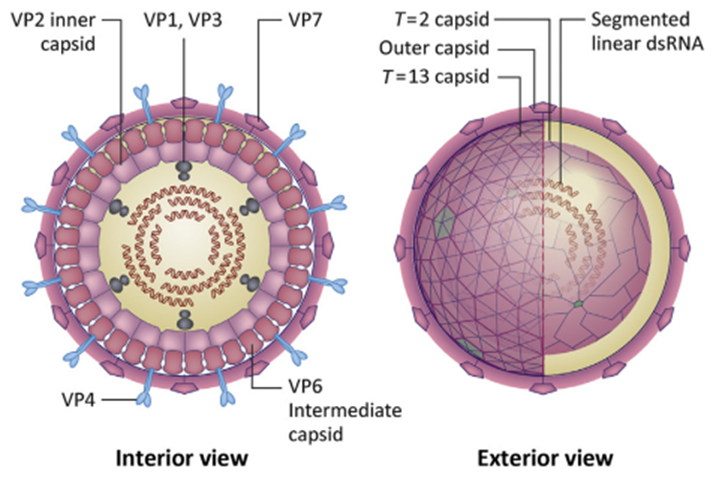
Figure 2. Structure of Reovirus Genome2. Reoviruses have segmented linear dsRNA that consist of VP1, VP2 (inner capsid), VP3, VP4, VP6 (middle capsid), and VP7 capsid. (VP: Viral protein)
Although virus particles of the family Reoviridae members have icosahedral symmetry, they might also be spherical, and the protein capsid surrounding linear dsRNA segments is organized as 60-80 nm wide, up to three concentric capsid protein layers2,6 (Figure3)2.
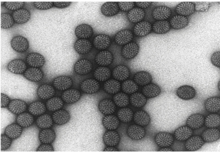
Figure 3. The Electron Micrograph Image of Triple-layered Rotavirus2. The negatively stained transmission electron microscopic (TEM) image facilitates observing some ultrastructural morphology of rotavirus particles.
Replication of Reoviruses
Orthoreoviruses are used as models to study Reovirus replication. Reovirus replication and transcription occur in the cytoplasm and capsids, respectively7,8. Virions attach to sialic acid receptors of the cell and enter the cytoplasm through endocytosis. After the entrance, the cores become transcriptionally active8,9. Negative RNA is first synthesized in the cytoplasm via primary and secondary transcription, followed by a viral protein synthesis7,9. Protein synthesis in the host cells declines since virions are attached to the cells8. Reovirus mRNAs have a 5′-terminal 7-methylguanosine (m7G) cap that allows translation into host cell ribosomes. However, they lack 3′ poly-A (poly- Adenine) tail found in highly translated cellular messages8,9. Reoviruses use an adhesion promoter mechanism to infect cells. This mechanism begins with the low-affinity interaction of σ1 attachment protein to the surface carbohydrates on the cell, followed by σ1 and junctional adhesion molecule-A (JAM-A) interaction1,10. In Orthoreoviruses, σ1 mediates the attachment to target cells. σ1 is a filamentous trimer seen as the spike on the virion. Varieties of σ1 define the cell and tissue tropism of viruses5-9. In viroplasm (viral factory), newly synthesized viral core proteins are associated with Reovirus mRNAs to produce new core particles. As a result, viral transcription boosts viral protein synthesis10,11. After attachment, Reovirus enters the cell via endocytosis in a β1 integrin-dependent manner9. In an endocytic way, acid-dependent cathepsin protease B and L removes σ3 and forms δ (delta) and φ, infectious subvirion particle (ISVP) entry intermediate by dividing µ1 into two pieces12. ISVP is also produced in the lungs and intestine by tissue-resident proteases during natural infection1,5,13. Φ fragments create pores in membranes. It is assumed that they function together with particle-associated δ fragments to mediate translocation of the viral core through the endosomal membrane and cytoplasm12,13 (Figure 4)9. Although exocytosis of Reoviruses is not fully understood, it is believed that it might occur through cell lysis4,14. Furthermore, the latest research suggests that Reoviruses are excreted from the cell via nonlytic mechanisms7. Some details of general replication steps in Reoviruses might vary from genera to genera. As an example, Rotaviruses and Arboviruses show differences in replication steps due to adaptation to different host environments8,9. The replication of Orbiviruses and Rotaviruses has similar properties to those of Orthoreovirus9. Rotaviruses require three-layered virus particles containing VP4 and VP7 for attachment. Proteolytic cleavage of VP4 (with chymotrypsin in the small intestine) is crucial to the entrance of viruses into cells and the rise of the infection13,14. Some Rotavirus strains attach to the sialic acid residues on the cell surface. Based on some assumptions, rotavirus may enter the cell via receptor-dependent endocytosis or direct penetration15.
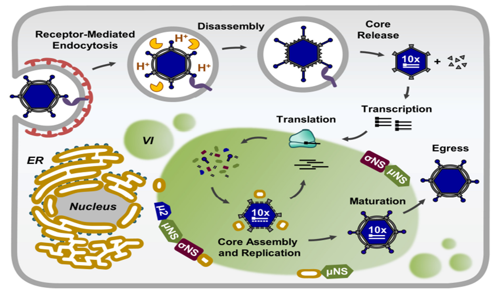
Figure 4. Reovirus Replication Cycle9. VI:viral inclusions; ER-endoplasmic reticulum.
Reverse Genetic Sytems
Reverse genetic systems allow the production of contagious viruses from cloned nucleic acids8. These systems are beneficial for understanding the functions of protein-coding and gene regulatory regions. When it comes to reverse genetic systems, not all Reoviruses are created equal8,15. As a result, systems based entirely on cloned genes have been created for some Orthoreovirus and Orbiviruses5,16. The mammalian Orthoreovirus system includes the transfection of cells with plasmids that encode and express every viral gene7. The Bluetongue virus (one kind of orbivirus) system has quite distinct features17. mRNAs are copied in vitro, and single-stranded RNAs (ssRNA) are transfected into the cells13. When a similar method was applied to rotaviruses, it did not succeed12,17. Since current Rotavirus reverse genetic systems rely on other helper viruses to infect helper viruses, their benefits are limited8. Rotaviruses are the most significant and common human pathogens in the family Reoviridae due to the diseases they cause9. Simple expression or integration of rotavirus mRNAs into the cell is insufficient to produce contagious viruses1.
Viral mRNA is synthesized in the core, and new transcripts are secreted via pores that present on the corners of the inner capsid assuming every genome segment is associated with its own pore and transcription complex1,2. mRNAs are not polyadenylated but form a hairpin structure at the 3′ end10. Viral transcript synthesis of rotaviruses is mediated by VP1 and VP3 complexes18. While VP1 contains the catalytic domain of RNA synthesis, VP3 functions as nucleotide phosphohydrolase, guanyl transferase, and methyltransferase as well as ssRNA binding17. For this reason, VP3 is known as the encapsulant component of the complex18. Transcriptionally active cores are extracted from purified virions (removal of the outer capsid layer). As a result, mRNA synthesis can reach up to milligrams with a sufficient number of in vitro premier sources17,18. A T=13 icosahedral capsid layer surrounds the core. Some Reoviruses (members of the Rotavirus family) have a third T=13 icosahedral protein layer12. Rotaviruses have glycosylated spike proteins2. In rotaviruses, non-structural protein 3 (NSP3) binds to eukaryotic initiation factor 4F (elF4F), a cap-binding complex, to facilitate translation12. Therefore, NSP3 helps host poly-A binding protein to function at the beginning of the translation19. Generally, rotavirus mRNAs are translated in the cytosol, but the mRNAs of the VP4, VP7, and NSP4 proteins are translated and glycosylated in the rough endoplasmic reticulum1,2,9. Virus factories, also known as viroplasm, are electron-dense areas of the cell and have organelle-like structures formed by cytosolic proteins. The genome replication and core assembly occur in the viroplasm. Cores are collected in the viroplasm and may be translated into more mRNAs1,4,9.
Epidemiology of Reoviruses
Reoviruses are common in human populations, but they are not associated with any human diseases20. However, rotaviruses, members of the Reovirus family, cause severe diarrhea in infants and constitute a major percentage of infant deaths in some regions9,12. Reoviruses are isolated from the respiratory and gastrointestinal tracts are called orphan viruses because their infections show no symptoms. 63% of 20–30-year-old adults and 10% of 1–5-year-old children in the US have antibodies against Reoviruses8.
Reoviruses are transmitted between animals directly or indirectly. Pig populations in the United States, Europe, and China are infected by Type 1-3 Reoviruses. Pigs become susceptible to infections after 11 weeks of immunity against Reoviruses12. The infection spreads by respiration, fecal-oral contact, or consuming contaminated food and water10,12. Reoviruses can survive in shelters and hatcheries because they are resistant to heat and disinfectants. Therefore, they are known as direct pathogens of some animal diseases8,15.
Reoviruses reproduce in the respiratory and gastrointestinal systems, but details about their pathophysiology are not available7. The S1 glycoprotein was considered crucial for cell attachment and entrance into respiratory epithelia at first. According to a recent study, S1 may not be enough for virus entrance into the cell, but it may play a role in the systemic spread of viruses and viremia1,2.
The virus can infect pigs’ gastrointestinal tracts, nervous systems, and leukocytes when they are infected through respiration12. Reoviruses are found in nasal secretions and feces 24 hours after vaccination, and shedding lasts 6–14 days21. Hemagglutination-inhibiting antibodies can be detected seven days after infection and peak between 11 and 21 days12,20,21.
Rotaviruses
Rotaviruses, which belong to the family Reoviridae, are unenveloped and cube-shaped viruses. They are 60-80 nm in size and have dsRNA as genetic material9,12,22. Virion structure consists of one or two-layered outer protein layers and one inner protein layer1,2. Members of the family Reoviridae can reproduce in cell culture and are resistant to environmental factors. They can survive at 70oC and show resistance to lipid dissolvers2,6,12. Rotaviruses are extremely infectious. Even water-born infections are well-documented, and transmission generally occurs via direct contact12,21.
Replacement of fluids and electrolytes is the primary treatment for rotavirus-induced dehydration21. The treatment involves intravenous fluid injections or oral fluid replacement therapy10. Rotaviruses cause gastrointestinal infections in humans and other mammals12,21.
The Rotavirus genome consists of six structural segments (VP1, VP2, VP3, VP4, VP6, VP7), each encodes for one protein, and five non-structural segments (NSP1-NSP5). These segments are divided into seven antigenic groups (A, B, C, D, E, F, and G)10,12,21. The antigenic groups and host organisms are shown in Table 212.
Table 2. Rotavirus groups and host organisms they infect12.
| GROUPS | HOST |
| A | Human, cattle, horse, pig, dog, cat, rabbit, mouse, bird |
| B | Human, pig, cattle, sheep, rats |
| C | Human, pig, ferret, cattle |
| D | Chicken |
| E | Pig |
| F | Chicken |
| G | Chicken |
The primary region of rotavirus replication is the small intestine because virions infect non-dividing erythrocytes located at villi10,12. Rotavirus infections are cytopathic and result in the loss of intestinal epithelial cells and disruption in the absorption of nutrients caused by mild diarrhea21,22. The rotavirus non-structural protein 4 (NSP4) is considered the primary cause of severe diarrhea symptoms8,12. NSP4 is a multi-functional protein that plays a role in rotavirus replication12. While some NSP4s are found in the ER membrane, infected cells also release some. Released NSP4s may be found on apical and basolateral sides of polarised epithelial cells. NSP4 binds to basolateral integrin to trigger cellular calcium mobilization (ER calcium release), which results in chloride release through calcium-activated channels. Following chloride release, water and sodium release occur, resulting in severe diarrhea9,12,22. Newly developed rotavirus vaccinations may minimize infection-related morbidity and mortality. Rotavirus vaccines are live-attenuated vaccines produced by re-dosing. They are effective against severe diseases but cannot prevent the disease directly12,21,22.
Rotavirus infections destroy terminally differentiated enterocytes that cover villi ends, leading to intestinal malabsorption and digestion8,12. The neonatal intestine is more susceptible to rotavirus infections because of the slow epithelial turnover rate and abundance of terminally differentiated enterocytes in the mucosal epithelium8,10,12. These infections have severe effects on newborns, while adults experience only subclinical effects. Damaged villi get shortened and covered by immature and less differentiated epithelial cells migrated from crypts9. These cells secrete reduced levels of disaccharidases such as lactase, and glucose-dependent sodium transport occurs less1,2. Undigested lactose in milk promotes bacterial reproduction and shows a high osmotic effect. Both mechanisms increase the symptoms of diarrhea14.
Orbiviruses
Insects are representatives of the orbiviruses, a member of the family of Reoviruses. Bluetongue virus (BTV), Epizootic hemorrhagic disease virus (EHDV), and African horse sickness virus (AHSV) are animal pathogens of orbiviruses1,2,16. There are 22 different varieties of orbiviruses that infect insects and other animals18. Ticks, mosquitoes, sandflies, and biting midges transmit these viruses, which are also known as “arboviruses.” Orbiviruses have a broad host range among birds and other animals. They cause cytopathic infections in animal cells, except for insect cells18,23,24.
Culicoides midges transmit Epizootic hemorrhagic disease virus (EHDV), a type of orbivirus, EHDV affects wild ruminants, especially white-tailed deer in North America, and it is one of the most significant deer diseases in North America1,2,6,16. The disease is characterized by a sudden and extreme fever, malaise, extreme salivation, rapid pulse, increased respiratory rate, and bleeding. The deer dies 8–36 hours after symptoms are observed16,18,24.
The African horse sickness virus (AHSV) is specific to Africa and infects horses, mules, donkeys, and zebras. It is transmitted to animals by Culicoides bites. While it causes severe symptoms among donkeys and horses, zebras do not show symptoms. Therefore, zebras are known as natural reservoirs of the virus18,23.
Orthoreoviruses
Orthoreoviruses, a member of the Reovirus family, are transmitted fecally and orally and infect birds, mammals, and reptiles. These viruses are common and rarely cause serious illness. They are so ubiquitous in feces that their existence can be a fecal contamination sign1,2,25. They cause mild upper respiratory tract disease, gastroenteritis, and biliary atresia2,25. Reoviruses were found in dead crows examined by West Nile virus monitoring programs in the United States. Similar analyses are used for the serological diagnosis of bird Orthoreovirus infections besides immunofluorescence. In addition, viruses are isolated from bird cell cultures when serological methods are insufficient25.
Avian Reoviruses
Avian Reoviruses are resistant to ether, chloroform, low pH (pH =3), trypsin, and sodium deoxycholate1,5. Unlike Mammalian Reoviruses, Avian Reoviruses do not display hemagglutination and hemadsorption. They are produced in embryonated eggs and primary cell cultures8,9. Avian Reoviruses are transmitted vertically, and the vertical transmission reduces the hatching rates and substantially increases early chick deaths1,2. Avian Reovirus infections show two main syndromes.
1-Malabsorption syndrome
Malabsorption syndrome displays diarrhea, malnutrition, decreased rate of feather growth, weakness in legs, skeleton deformation, broiler growth arrest, runtiness, and low skin pigmentation2,24,26. Because of its many symptoms, this syndrome was characterized as a deficiency of minerals (Ca, P) and vitamins (A, B, D, E), viral infections (IBD), toxications (mycotoxicosis), and protozoan diseases (coccidiosis). However, Malabsorption syndrome is caused by Reoviruses, according to the latest studies4,5,26.
2-Viral arthritis/tenosynovitis
Viral arthritis/tenosynovitis is usually seen in chickens and turkeys. The disease is characterized by synovial membranes and myocardium (heart muscle) inflammation, arthritis (joint inflammation), and lameness. In acute cases, underdevelopment of flocks, malnutrition, and death occur5,26.
Coltiviruses
Coltiviruses are members of the subfamily Spinareovirinae, and they have a genome with 12 segments. Vertebrates, rodents, and ungulates (sheep and deer) are hosts of the Colorado tick fever virus (CTFV) in the US1,2. CTHV may cause flu-like symptoms, meningitis, encephalitis, and other severe symptoms1. First, CTFV was considered a mild Rocky Mountain spotted fever caused by Rickettsia rickettsii. However, CTFV is an arbovirus that spreads via infected tick bites18. Infections in humans are most common at elevations of 4000-10.000 feet in the spring and summer, when ticks are most active18,26.
Reoviral Therapy for Cancer
Orthoreovirus, mammalian Reovirus, is considered one of the oncolytic viruses developed as cancer therapeutics. Clinical findings of naturally occurring Reovirus infections make Reovirus an ideal candidate for cancer virotherapy in immunocompromised patients25,27,28. Many clinical phase 1 and phase 2 trials showed that T3 Dearing (T3D) strain-based Reovirus (Pelareorep) is safe for cancer patients undergoing immunosuppressive treatments13. Reovirus infections make tumor cells susceptible to chemotherapeutic drugs and stimulate cell-associated immunity. Therefore, Reovirus infections are considered a good therapeutic agent and a good candidate for combination therapy13,27,28. Epidermal growth factor receptor (EFGR) is stimulated by RAS, and this increased stimulation may result in increased proliferation and survival rate of tumor cells3. RAS stimulation causes increased Reovirus replication, particle generation, and apoptosis-mediated release of projectile virions. Proteolytic degradation during the entry of Reovirus virion into the cells and detection of susceptibility to Reoviruses are crucial in terms of Reovirus oncolysis13,25. Although RAS has a major effect on Reovirus oncolysis, tumor cells may be killed via RAS-independent mechanisms3. For example, the destruction of head and neck cancer and lung cancer cell lines via Reoviruses does not indicate RAS transformation. Reovirus infection in most cancer cells is associated with the cell surface receptor Junctional Adhesion Molecule A (JAM-A), which plays a role in the entry of Reovirus into the cells26,27. However, JAM-A expression is not essential for tumor cell infection in all cancer types. For example, glioblastoma infection with Reoviruses depends on JAM-A in 2D cultures, while it occurs independently of JAM-A in 3D cultures3,26,27. These findings suggest that cellular microenvironments influence Reovirus oncolysis susceptibility3.
Reoviruses and Celiac disease
Celiac disease (CD) causes autoimmune enteropathy in individuals genetically sensitive to gluten consumption9,27. One out of every 133 people in America has CD, but most are undiagnosed28. Children with CD suffer from diarrhea and malabsorption syndrome. In addition, the CD could cause extraintestinal autoimmune disorders, infertility, miscarriage, and cancer9,27,28. Gluten consumption is the most significant factor related to CD. Therefore, current treatment strategies stress gluten-free diets for people with CD10,29. CD pathogenesis must be well understood due to its increased prevalence and the consequences of misdiagnosis3,27,29.
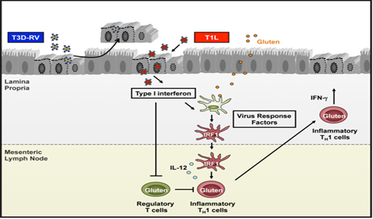
Figure 5. Celiac Disease Mechanism29. Reovirus T1L and T3D-RV infect the intestine after peroral inoculation. T3D-RV activates caspase-3 and leads to sloughing of intestinal epithelial cells, resulting in rapid viral clearance. Nevertheless, T1L subverts the antiviral response to prolong infection by releasing type 1 interferons and other virus-response factors (yet to be identified) that cause IRF-1 to be expressed in dendritic cells in the lamina propria. Dendritic cells enter the mesenteric lymph nodes and release IL-12 that activates gluten-specific inflammatory T cells (TH1) in response to food antigens such as gluten.The type 1 interferons produced during T1L infection inhibit regulatory T cells, which leads to the development of TH1 immunity to gluten during celiac disease development. IRF-1:interferon regulatory factor-1; TH1: gluten-specific inflammatory T cells
Human Orthoreovirus (Reovirus) strains can infect rats orally, activating innate immune pathways similar to rotavirus1,2,5. Reovirus also causes type 1 IFNs production. When rats are fed the model antigen ovalbumin (OVA), they develop systemic tolerance to tagged OVA, with Treg activation and the absence of OVA-specific inflammatory TH1 cells9,10. HLA-DQ8 ± transgenic rats inoculated with T1L and fed with gliadin, a proteolytic derivative of gluten, develop gluten-specific antibodies and DTH response against gluten, suggesting rats do not have gluten tolerance28. T1L infection activates transglutaminase 2, an enzyme that enhances CD immunopathogenesis. In this way, Reovirus inflammation that leads to virus-mediated CD is triggered28,29.
Reovirus infections are common in early childhood in humans when maternal immunity reduces and diets begin to contain solid foods, including wheat flakes27,28. In this period, children are the most susceptible to CD. CD patients exhibit higher levels of Reovirus antibodies than control people, suggesting that Reovirus infections may contribute to CD development3,29. In one study, rotavirus, another family member of the Reoviridae, was associated with CD onset but not in later studies. The inconsistency of findings implies the importance of researching clinical and mechanical phenomena to understand the relationship between viral infections and CD3,28,29.
Conclusion
Reoviruses are common viruses that infect a broad range of strains due to their high variations. They cause severe illnesses in animals worldwide and are mainly responsible for infant deaths in humans. On the other hand, Reoviruses are useful for reoviral cancer therapy since they are oncolytic viruses2,5,9,10.
Acknowledgment
Finally, we would like to thank the author and the editor of the Turkish version for their contributions. The review article is available in Turkish on www.bioinforange.com.
References
1.Reoviridae. (2012). In Virus Taxonomy (pp. 541–637). Elsevier. https://doi.org/10.1016/b978-0-12-384684-6.00051-3
2.Payne, S. (2017). Family Reoviridae. In Viruses (pp. 219–226). Elsevier. https://doi.org/10.1016/b978-0-12-803109-4.00026-x
3.Zhao, X., Chester, C., Rajasekaran, N., He, Z., & Kohrt, H. E. (2016). Strategic combinations: The future of oncolytic virotherapy with reovirus. In Molecular Cancer Therapeutics (Vol. 15, Issue 5, pp. 767–773). American Association for Cancer Research Inc. https://doi.org/10.1158/1535-7163.MCT-15-0695
4.Kibenge, F. S. B., & Godoy, M. G. (2016). Reoviruses of Aquatic Organisms. In Aquaculture Virology (pp. 205–236). Elsevier Inc. https://doi.org/10.1016/B978-0-12-801573-5.00014-0
5.Outline, C. (2017). Reoviridae. In Fenner’s Veterinary Virology (pp. 299–317). Elsevier. https://doi.org/10.1016/b978-0-12-800946-8.00015-5
6.Strauss, J. H., & Strauss, E. G. (2008). Viruses That Contain Double-Stranded RNA: Family Reoviridae (2nd ed., pp. 193–210). https://doi.org/10.1016/b978-0-12-373741-0.50008-8
7.Roth, A. N., Aravamudhan, P., Fernández de Castro, I., Tenorio, R., Risco, C., & Dermody, T. S. (2021). Ins and Outs of Reovirus: Vesicular Trafficking in Viral Entry and Egress. In Trends in Microbiology (Vol. 29, Issue 4, pp. 363–375). Elsevier Ltd. https://doi.org/10.1016/j.tim.2020.09.004
8.Kanai, Y., & Kobayashi, T. (2021). Annual Review of Virology FAST Proteins: Development and Use of Reverse Genetics Systems for Reoviridae Viruses. https://doi.org/10.1146/annurev-virology-091919
9.Tenorio, R., de Castro, I. F., Knowlton, J. J., Zamora, P. F., Sutherland, D. M., Risco, C., & Dermody, T. S. (2019). Function, architecture, and biogenesis of reovirus replication neoorganelles. In Viruses (Vol. 11, Issue 3). MDPI AG. https://doi.org/10.3390/v11030288
10.Bouziat, R., Hinterleitner, R., Brown, J. J., Stencel-Baerenwald, J. E., Ikizler, M., Mayassi, T., Meisel, M., Kim, S. M., Discepolo, V., Pruijssers, A. J., Ernest, J. D., Iskarpatyoti, J. A., Costes, L. M. M., Lawrence, I., Palanski, B. A., Varma, M., Zurenski, M. A., Khomandiak, S., Mcallister, N., … Xavier, R. J. (9 C.E.). Reovirus infection triggers inflammatory responses to dietary antigens and development of celiac disease (Vol. 9). https://doi.org/10.1146/annurev-virology-091919-070225
11.Dallari, S., Heaney, T., Rosas-Villegas, A., Neil, J. A., Wong, S. Y., Brown, J. J., Urbanek, K., Herrmann, C., Depledge, D. P., Dermody, T. S., & Cadwell, K. (2021). Enteric viruses evoke broad host immune responses resembling those elicited by the bacterial microbiome. Cell Host and Microbe, 29(6), 1014-1029.e8. https://doi.org/10.1016/j.chom.2021.03.015
12.Shepherd, F. K., Freeman, M. J., Culhane, M. R., & Marthaler, D. G. (2019). Reoviruses (rotaviruses and reoviruses). Diseases of Swine (11th ed.). https://doi.org/10.1002/9781119350927.ch43
13.Comins, C., Simpson, G. R., Relph, K., Harrington, K. J., Melcher, A., & Pandha, H. (2013). Reoviral Therapy for Cancer: Strategies for Improving Antitumor Efficacy Using Radio- and Chemotherapy. In Gene Therapy of Cancer: Translational Approaches from Preclinical Studies to Clinical Implementation: Third Edition (pp. 185–198). Elsevier Inc. https://doi.org/10.1016/B978-0-12-394295-1.00013-5
14.McNamara, A., Roebke, K., & Danthi, P. (2020). Cell Killing by Reovirus: Mechanisms and Consequences. https://doi.org/10.1007/82_2020_225
15.Zhang, X., Lei, X., Ma, L., Wu, J., & Bao, E. (2019). Genetic and pathogenic characteristics of newly emerging avian reovirus from infected chickens with clinical arthritis in China. Poultry Science, 98(11), 5321–5329. https://doi.org/10.3382/ps/pez319
16.Cottingham, S. L., White, Z. S., Wisely, S. M., & Campos-Krauer, J. M. (2021). A mortality-based description of ehdv and btv prevalence in farmed white-tailed deer (Odocoileus virginianus) in florida, USA. Viruses, 13(8). https://doi.org/10.3390/v13081443
17.Mertens, P., Mohd Jaafar, F., & Attoui, H. (2015). Orbiviruses and Bluetongue Virus. In eLS (pp. 1–18). Wiley. https://doi.org/10.1002/9780470015902.a0001010.pub3
18.Song, S., Li, Y., Fu, S., Lei, W., Guo, X., Feng, Y., Gao, X., Li, X., Yang, Z., Xu, Z., He, Y., Wang, H., Zhou, H., Wang, B., Lu, X., & Liang, G. (2017). Genome sequencing and phylogenetic analysis of Banna virus (genus Seadornavirus, family Reoviridae) isolated from Culicoides. Science China Life Sciences, 60(12), 1372–1382. https://doi.org/10.1007/s11427-017-9190-6
19.Egaña-Labrin, S., Hauck, R., Figueroa, A., Stoute, S., Shivaprasad, H. L., Crispo, M., Corsiglia, C., Zhou, H., Kern, C., Crossley, B., & Gallardo, R. A. (2019). Genotypic Characterization of Emerging Avian Reovirus Genetic Variants in California. Scientific Reports, 9(1). https://doi.org/10.1038/s41598-019-45494-4
20.Koehler, M., Petitjean, S. J. L., Yang, J., Aravamudhan, P., Somoulay, X., lo Giudice, C., Poncin, M. A., Dumitru, A. C., Dermody, T. S., & Alsteens, D. (2021). Reovirus directly engages integrin to recruit clathrin for entry into host cells. Nature Communications, 12(1). https://doi.org/10.1038/s41467-021-22380-0
21.Stettner, E., Dietrich, M. H., Reiss, K., Dermody, T. S., & Stehle, T. (2015). Structure of Serotype 1 Reovirus Attachment Protein σ1 in Complex with Junctional Adhesion Molecule A Reveals a Conserved Serotype-Independent Binding Epitope. Journal of Virology, 89(11), 6136–6140. https://doi.org/10.1128/jvi.00433-15
22.Long, C. P., & McDonald, S. M. (2017). Rotavirus genome replication: Some assembly required. In PLoS Pathogens (Vol. 13, Issue 4). Public Library of Science. https://doi.org/10.1371/journal.ppat.1006242
23.Zientara, S., Weyer, C. T., & Lecollinet, S. (n.d.-w). African horse sickness. OIE Revue Scientifique et Technique, 34(2), 315–327. https://doi.org/10.20506/rst.34.2.2359
24.Belbis, G., Zientara, S., Bréard, E., Sailleau, C., Caignard, G., Vitour, D., & Attoui, H. (2017). Bluetongue Virus: From BTV-1 to BTV-27. In Advances in Virus Research (Vol. 99, pp. 161–197). Academic Press Inc. https://doi.org/10.1016/bs.aivir.2017.08.003
25.Phillips, M., Stuart, J., Rodríguez Stewart, R., Berry, J., Mainou, B., & Boehme, K. (2018). Current understanding of reovirus oncolysis mechanisms. Oncolytic Virotherapy, Volume 7, 53–63. https://doi.org/10.2147/ov.s143808
26.Dietrich, M. H., Ogden, K. M., Long, J. M., Ebenhoch, R., Thor, A., Dermody, T. S., & Stehle, T. (2018). Structural and Functional Features of the Reovirus σ1 Tail. Journal of Virology, 92(14). https://doi.org/10.1128/jvi.00336-18
27.Eledge, M. R., Zita, M. D., & Boehme, K. W. (2019). Reovirus: Friend and Foe. In Current Clinical Microbiology Reports (Vol. 6, Issue 3, pp. 132–138). Springer. https://doi.org/10.1007/s40588-019-00121-8
28.Giovanna, Z., Marzia, D., Claudio, L., & Puccetti, A. (2015). Celiac Disease and Rotavirus Infection. In Infection and Autoimmunity (pp. 453–464). Elsevier Inc. https://doi.org/10.1016/B978-0-444-63269-2.00028-3
29.Brown, J. J., Jabri, B., & Dermody, T. S. (2018). A viral trigger for celiac disease. In PLoS Pathogens (Vol. 14, Issue 9). Public Library of Science. https://doi.org/10.1371/journal.ppat.1007181