
Reliable analysis of biological data obtained through microscopy is largely dependent on accurate cell segmentation. Thanks to new-generation deep learning (DL) technologies, two-dimensional (2D) cell segmentation offers generalised solutions across various cell types and imaging methods. This success stems from the relatively easy scalability of image acquisition, labelling, and computation. However, three-dimensional (3D) cell segmentation still poses a significant challenge due to the requirement for densely labelled 2D slices. Manually labelling 3D cells is a time-consuming, uncertain, and almost impractical process for training large-scale models.
Zhou and his team have developed u-Segment3D, a new toolset that facilitates the transition from 2D segmentations to 3D composite segmentations and does not require training data. The research team demonstrated u-Segment3D’s effectiveness on real-life datasets, from single cells to cell clusters and tissues, by showcasing its ability to convert and improve 2D-to-3D segmentation, making it compatible with pixel-based sample cell masks generated by existing 2D methods. This approach proved that u-Segment3D is more effective than 3D segmentations when cells are crowded or have complex morphologies.
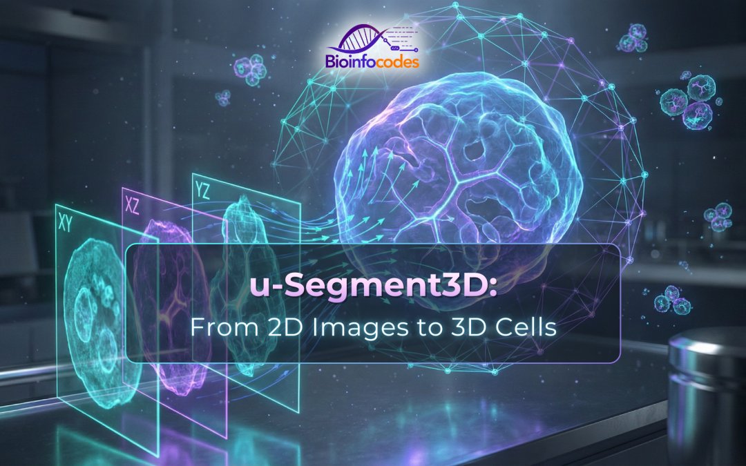
u-Segment3D formulates obtaining 3D segmentations from 2D segmentations as a general optimisation problem. Researchers have reconstructed 3D gradient vectors for each cell based on its 3D medial axis and the corresponding distance transform representation. In this process, using gradient descent and spatially connected component analysis, a consistent 3D fused segmentation was obtained from 2D segmentations. The method is designed to generate a consensus 3D segmentation from 2D segmentation outputs in a stack of one, two, or three orthoviews. The entire process worked independently of cell morphology and the 2D segmentation method used, providing a universal solution. u-Segment3D has been validated on 11 different real-world datasets containing over 70,000 cells, ranging from single cells to cell clusters, embryos to tissues, and entire vascular networks.
In conclusion, the findings demonstrate that u-Segment3D exhibits performance comparable to, and often surpassing, established 3D segmentation methods, even in environments with complex cell morphologies and dense cell populations. This progress is of great importance, especially for microscope-based biological research, where labelling large and diverse 3D cell datasets is difficult. Thanks to its approach that doesn’t require training data, u-Segment3D offers significant potential in this field. It is anticipated that future studies will continue to investigate the effects of this technology on broader biological processes and different 3D imaging techniques.
Reference: Zhou, F.Y., Marin, Z., Yapp, C. et al. Universal consensus 3D segmentation of cells from 2D segmented stacks. Nat Methods 22, 2386–2399 (2025). https://doi.org/10.1038/s41592-025-02887-w