

Ece GüvenA., Elif DuymazE., Sema YöşiliR.
Introduction
After the discovery of clustered regularly interspaced short palindromic repeats (CRISPR)-associated (Cas) system in bacterial adaptive immunity against foreign genetic elements like viruses, they have been frequently used to edit genomes in a much easier way compared to previously utilized methods such as transcription activator-like effector nucleases (TALENs) and zinc finger nuclease (ZFNs). The reason is that CRISPR-Cas does not need protein engineering to edit the genome, unlike TALENs or ZFNs. This system can be adapted for applications of diagnostics and gene therapy. Many promising trials have tested the use of CRISPR-Cas in correcting mutations associated with various diseases including rare and hereditary ones such as hemophilia, β-thalassemia, Duchenne muscular dystrophy, cystic fibrosis, Huntington’s and Alzheimer’s disease1.
With the advancements in the CRISPR-Cas-based editing technologies, there have been many versions of the initially utilized system for improving efficiency, accuracy, and safety in clinical applications. The tools can insert and delete a gene, as well as make base pair (bp) conversions to correct the diseases2. In this review, four different tools of CRISPR-Cas application in gene editing technologies, which are double-stranded DNA breakage (DSB) repair pathways, base editing, prime editing, and CRISPR-associated transposase will be evaluated, each having its advantages and disadvantages.
CRISPR-Cas Mechanism
The CRISPR-Cas system works by complex formation of Cas proteins with the CRISPR RNAs (crRNA or guide RNA), which are encoded by CRISPR arrays. The crRNA guides the complex to recognize the target sequence, generally through protospacer adjacent motif (PAM) or protospacer flanking sequence (PFS) on foreign genetic elements, allowing Cas proteins to cleave the target strands by their nuclease activities3. The system was first discovered by Mojica, who found CRISPR loci in many microbes that construct adaptive immunity of the microorganisms against specific infections. Then, Charpentier and Doudna in their study, revealed that Cas9 of Streptococcus pyogenes can be programmed to a target location with an engineered guide RNA (gRNA) to cleave the double stranded DNA (dsDNA) in vitro, as a novel genome editing application4-6.
CRISPR-Cas systems could be classified into two groups, with respect to the construction of effector proteins, as Class 1 and Class 2. A majority of Cas proteins which are utilized in gene editing applications such as Cas9, 12, 13, and 14, are involved in Class 2 since they are easier to utilize due to incorporation of only one effector protein. On the other hand, many effector proteins build up a complex in the Class 1 system. Some of these Cas proteins may need a transactivating RNA (tracrRNA) for cleavage of the target or crRNA maturation. They may cleave double or single-stranded DNA or RNA depending on the type of Cas proteins7,8.
Editing via Double Strand Breakage (DSB) Repair
The initial applications of the CRISPR-Cas system were based on dsDNA cleavage to trigger DNA repair pathways in dividing cells, including homology directed repair (HDR) and error prone non-homologous end joining (NHEJ) with the use of Class 2 proteins; especially Cas9 and single guide RNAs (sgRNA). With the HDR mechanism of the cell, large insertions can be achieved at a targeted location by adding a donor DNA having homology arms to the target sequence for correction of the gene of interest. On the other hand, knockouts can be performed with the NHEJ mechanism via causing insertion and deletion (indel) mutations (Figure 1)9.
As NHEJ is an error prone and random event that leads to off-target effects, mostly HDR mechanism is utilized for gene therapy. NHEJ mechanism predominates and occurs very frequently in a dividing cell, whereas the HDR mechanism is limited to the S/G2 phase of the cell cycle. Therefore, major disadvantages of this strategy are the inefficiency of the editing due to dependence on the host cell’s infrequent HDR machinery, and safety problems caused by Cas9’s potential to make on-target mutagenesis including complex genomic rearrangements and large deletions10-12. For these reasons, scientists have been developing more specific, efficient and safe ways for genome editing without creating a DSB.
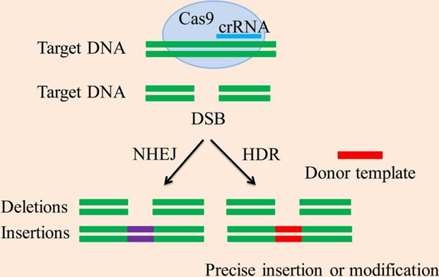
Figure 1. DSB repair based CRISPR gene editing via NHEJ and HDR mechanisms9. While NHEJ creates indel mutations in the target genome, HDR leads to the insertion of the desired sequence into the precise location in the genome. DSB: double-stranded DNA break, NHEJ: non-homologous end joining, HDR: homology directed repair.
Base Editing
Base editing has been utilized for site-specific modification in case of single nucleotide polymorphism (SNP) associated diseases, which does not create DSB in the genome so that mutations at the target site as well as off-target effects can be minimized. Base editors are composed of gRNA guided catalytically inactive Cas9 (dead, dCas9) or Cas9 nickase (nCas9) fused with cytidine deaminases (CBEs) or adenine deaminases (ABEs) that allow the conversion of cytidine (C) to thymidine (T) or adenine (A) to guanine (G) at the target site, respectively13. There are several base editors designed by scientists, which can be chosen for a study according to the purpose and cell types. Also, novel base editors have been engineered, and they collectively enable all the four transitions, as well as C to G transversion with different editing window preferences (Figure 2)13-15.
Liu et al. developed the first generation of CBE (BE1) where they fused a rat-derived cytosine deaminase Apolipoprotein B mRNA Editing Enzyme Catalytic Subunit 1 (APOBEC1) with dCas9. First uracil (U) is produced through hydrolytic deamination by CBEs so that uracil DNA glycosylases (UDG) can transform it to an apurinic/apyrimidinic (AP) site, activating the cell’s base excision repair pathway with the potential to create indels. Therefore, improved versions of CBE, called BE2 and BE3, have been developed by the addition of UDG inhibitor (UGI) to the BE1 and replacement of dCas9 with nCas9 in the BE2 system, respectively, for preventing dependence on cellular repair machinery. While BE2 offers editing via very little indel formation (~0.1%), BE3 can be utilized for more efficient editing with a drawback of increased indel formation (~1%)16,17. In theory, ABEs can correct half of the pathogenic mutations by converting A-T bp to G-C. Liu and his colleagues also engineered an adenosine deaminase, which does not occur naturally as cytidine deaminases, called ecTadA from Escherichia coli (E. coli) tRNA adenosine deaminase that converts A to inosine (I). ABE1.2 was the first generation ABE and was developed by the fusion of ecTadA variant, TadA*, with nCas9. Unlike CBEs, the ABE did not create any indel and off-target effects. After several experiments with protein engineering, they could create seventh generation ABE, ABE7.10, increasing the efficiency to 50% in human cells14,18. The major drawback of ABEs was their incompatibility with Cas9 orthologs other than SpCas9. Therefore another research group improved them into generation eight, ABE8e, which further advances efficiency and overcomes the compatibility issue with various Cas9 orthologs19.
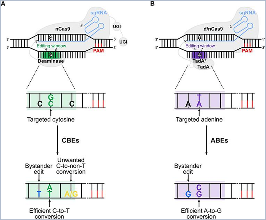
Figure 2. Cytosine base editors (CBEs) and adenine base editors (ABEs)20. A) CBEs are composed of cytidine deaminases fused with single gRNA (sgRNA) guided nCas9 and UGI for C-G conversion to T-A within editing window between nucleotide 4 and 8 (green). While BE3 has one UGI that is bound to the C terminus of the editor, BE4 has two UGIs. B) sgRNA guided dead (d) or nickase (n) Cas9 (d/nCas9) can be bound to two TadAs in ABEs, one being the evolved variant to edit A in DNA (TadA*) and the other being wild type (TadA). ABEs enable the conversion of A-T into G-C in the editing window between nucleotides 4 and 8 (purple). The off-target effect is shown in yellow in A and the bystander edit is shown in blue.
Bystander edits can be created by all base editors due to the presence of multiple C or A in the editing window. However, it generally does not occur or leads to silent mutations because of the degeneracy in genetic code or conservative mutations so that amino acid changes will not cause any difference. For other cases where bystander editing is problematic, the researchers can narrow the editing window or use high context dependent base editors for minimizing the bystander effect21.
Prime Editing
Recently, prime editing has emerged as a precise and effective genome editing tool with minimum indel formation, developed by Anzalone et al. Besides not relying on DSB or donor template, prime editing enables all possible single bp conversions, deletions up to 80 bp and insertions up to 44 bp at the target site. The target sites can be up to more than 30 bp away from the binding site, while a PAM sequence adjacent to the target site may not be needed20,22.
The prime editing includes one plasmid encoding for nCas9 that is conjugated with reverse transcriptase (RT) enzyme of (together called PE) and second plasmid encoding for prime editing guide RNA (pegRNA). The pegRNA is composed of a spacer sequence that is complementary to the target DNA, the primer binding site (PBS) for reverse transcription and the desired edit sequence at the 3’ end. The PE-pegRNA complex binds to one strand of the target DNA and nicks the opposite strand after recognition of the target site by the spacer in the pegRNA. Then, PBS is hybridized with the exposed DNA at the 3’ end so that the edit sequence in the pegRNA is reverse transcribed, enabling incorporation of the edit to the targeted site in the genome (Figure 3)2,23.PE1 was the first prime editor developed where wild-type Moloney murine leukemia virus (M-MLV) RT is used for fusion with nCas9. After the hypothesis that mutant RT can reveal enhanced binding to the template:PBS complex, enzyme processivity and thermostability, the researchers improved the version to PE2 by constructing a pentamutant RT which allowed about a 5 fold increase in editing efficiency. Moreover, the PE3 system has been evolved by adding sgRNA into the PE2 for nicking the non-edited strand as well to further enhance efficiency, with a small increase in indels and optimal nicking position being dependent on the target site22.
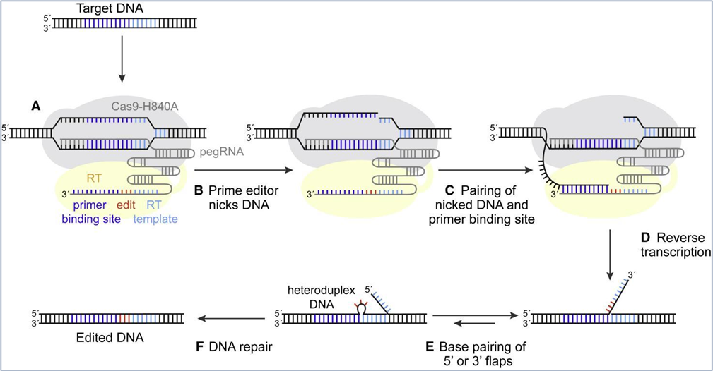
Figure 3. Principle of prime editing24. The description is given in the text above. After reverse transcription of the edit sequence, the edit sequence competes with the 5’ flap sequence for attaching to the target site. Then, 5’ flap is excised, and the edit sequence is ligated. Finally, DNA repair replaces the complementary strand by using the edited strand as a template. Cas9-H840A: Cas9 nickase (nCas9); RT: reverse transcriptase; pegRNA: prime editing guide RNA.
CRISPR-associated Transposase System
Many CRISPR-Cas systems that do not have nuclease activity have been identified until now, and an association of them with Tn7-like transposases was found by genomic and phylogenetic analysis25. In 2019, the study of Zheng et al. was the first to report the functional data of this association and propose a CRISPR-associated transposase system as an alternative targeted DNA integration tool. In their study, they characterized the natural CRISPR-associated transposases (CASTs) of cyanobacteria Scytonema hofmanni (ShCAST)to overcome limitations of DSB repair pathways and base editing which enables only nucleotide substitutions. ShCAST consists of Tn7-like transposase subunits and Cas12k which is a type V-K CRISPR effector with inactivated nuclease domain. They hypothesized that transposons may hijack the Cas effectors to enhance spread via phages or plasmids by generating R-loops at the target locus. Indeed, the results supported that Tn7-like transposases can be assisted by gRNAs to target sites for transposition of the transposon. The system could achieve 60% and 80% integration efficiency in the E. coli genome26.
The ShCAST system is mediated by the interaction of Cas12k with tnsC, tnsB and tniQ subunits after recognition of the target by sgRNA, allowing DNA integration at 49–66 bp downstream of G- and T-rich PAM. Another method has been developed called INTEGRATE (INsert Transposable Elements by Guide RNA-Assisted TargEting)which is a type I transposon-associated CRISPR system and uses an RNA guided cascade complex encoded by Vibrio cholerae Tn6677 transposon, composed of Cas6,7,8 proteins for DNA binding. The complex then interacts with the subunits tnsA, tnsB, tnsC and tniQ for integration of the cargo gene into the target site downstream of 46-55 bp of C-rich PAM. The transposon subunits recognize the left end (LE) and right end (RE) flanked on the cargo gene and excise the cargo at the donor. Then, they insert the cargo into the target DNA while creating gaps at both 5’ junctions which are repaired to make 5 bp target site duplication (Figure 4)13,21,27,28.
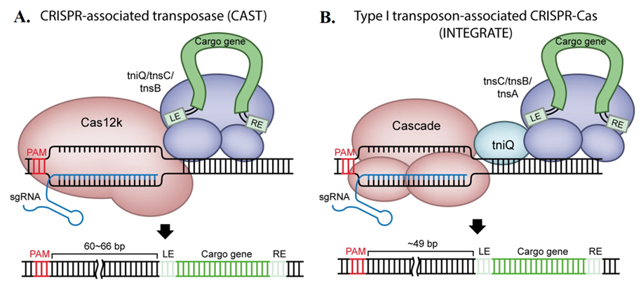
Figure 4. Description of the two CRISPR-associated transposase (CAST) methods28. A) ShCAST complex is directed to the target with sgRNA guided Cas12k and inserts the cargo DNA by its transposase subunits tniQ, tnsC and tnsB. B) INTEGRATE system includes three Cas proteins constructing the cascade which then forms a complex with tniQ and other transposase subunits to trigger DNA integration into the target genome.
The transpose-associated CRISPR methods offer safer and more specific and integration of DNA, up to 10 kb in length, compared to DSB repair-based genomic manipulations. However, the system has not been studied on mammalian cells or animal models and there is not much data for off-target effects. Also, the need for a specific PAM sequence further limits the use of the method. Therefore, the method should be improved to broaden its applications and should be tested for safety and efficiency in mammalian systems2,28.
References:
1.Mani, I. (2021). CRISPR-Cas9 for treating hereditary diseases: Reprogramming the Genome: CRISPR-Cas-Based Human Disease Therapy. Progress in Molecular Biology and Translational Science, 181, 165–183. https://doi.org/10.1016/bs.pmbts.2021.01.017
2. Domm, J. M., Wootton, S. K., Medin, J. A., & West, M. L. (2021). Gene therapy for Fabry disease: Progress, challenges, and outlooks on gene-editing. Molecular Genetics and Metabolism, 134(2), 117-131. https://doi.org/10.1016/j.ymgme.2021.07.006
3. Pickar-Oliver, A., Gersbach, C.A. (2019). The next generation of CRISPR–Cas technologies and applications. Nat Rev Mol Cell Biol 20, 490–507. https://doi.org/10.1038/s41580-019-0131-5
4. Lander, E. S. (2016). The Heroes of CRISPR. Cell, 164(1-2), 18.28. https://doi.org/10.1016/j.cell.2015.12.041
5. Mojica, F.J., Díez-Villaseñor, C., García-Martínez, J., Soria, E. (2005). Intervening Sequences of Regularly Spaced Prokaryotic Repeats Derive from Foreign Genetic Elements. J Mol Evol, 60, 174–182. https://doi.org/10.1007/s00239-004-0046-3
6. Jinek, M., Chylinski, K., Fonfara, I, Hauer, M., Doudna, J. A., Charpentier, E. (2012). A programmable dual-RNA-guided DNA endonuclease in adaptive bacterial immunity. Science, 337, 816-821. https://doi.org/10.1126/science.1225829
7. Kaminski, M. M., Abudayyeh, O.O., Gootenberg, J.S., Zhang, F., Collins, J. J. (2021). CRISPR-based diagnostics. Nat Biomed Eng, 5, 643–656. https://doi.org/10.1038/s41551-021-00760-7
8. Koonin, E. V., Makarova, K. S., Zhang, F. (2017). Diversity, classification and evolution of CRISPR-Cas systems. Current Opinion in Microbiology, 37, 67–78. https://doi.org/10.1016/j.mib.2017.05.008
9. Tang, X-D., Gao, F., Liu, M-J., Fan, Q-L., Chen, D-K., Ma, W-T. (2019). Methods for Enhancing Clustered Regularly Interspaced Short Palindromic Repeats/Cas9-Mediated Homology-Directed Repair Efficiency. Front. Genet., 10(551). https://doi.org/10.3389/fgene.2019.00551
10. Kosicki, M., Tomberg, K., & Bradley, A. (2018). Repair of double-strand breaks induced by CRISPR–Cas9 leads to large deletions and complex rearrangements. Nature Biotechnology, 36, 765-771. https://doi.org/10.1038/nbt.4192
11. Arnoult, N., Correia, A., Ma, J., Merlo, A., Garcia-Gomez, S., Maric, M., … Karlseder, J. (2017). Regulation of DNA repair pathway choice in S and G2 phases by the NHEJ inhibitor CYREN. Nature, 549(7673), 548–552. https://doi.org/10.1038/nature24023
12. Ray, U., & Raghavan, S. C. (2020). Modulation of DNA double-strand break repair as a strategy to improve precise genome editing. Oncogene, 39, 6393–6405. https://doi.org/10.1038/s41388-020-01445-2
13. Moon, S. B., Kim, D. Y., Ko, JH., Kim, YS. (2019). Recent advances in the CRISPR genome editing tool set. Exp Mol Med, 51, 1–11. https://doi.org/10.1038/s12276-019-0339-7
14. Kantor, A., McClements, M. E., MacLaren, R. E. (2020). CRISPR-Cas9 DNA Base-Editing and Prime-Editing. International Journal of Molecular Sciences, 21(17). https://doi.org/10.3390/ijms21176240
15. Kurt, I.C., Zhou, R., Iyer, S. et al. (2021). CRISPR C-to-G base editors for inducing targeted DNA transversions in human cells. Nat Biotechnol, 39, 41–46. https://doi.org/10.1038/s41587-020-0609-x
16. Komor, A., Kim, Y., Packer, M. S., Zuris, J. A., Liu, D. R. (2016). Programmable editing of a target base in genomic DNA without double-stranded DNA cleavage. Nature 533, 420–424. https://doi.org/10.1038/nature17946
17. Wang, L., Xue, W., Yan, L. et al. Enhanced base editing by co-expression of free uracil DNA glycosylase inhibitor. Cell Res 27, 1289–1292 (2017). https://doi.org/10.1038/cr.2017.111
18. Gaudelli, N. M., Komor, A. C., Rees, H. A., Packer, M. S., Badran, A. H., Bryson, D. I., Liu, D. R. (2017). Programmable base editing of A•T to G•C in genomic DNA without DNA cleavage. Nature, 551, 464-471. https://doi.org/10.1038/nature24644
19. Richter, M.F., Zhao, K.T., Eton, E., Liu, D. R., et al. (2020). Phage-assisted evolution of an adenine base editor with improved Cas domain compatibility and activity. Nat Biotechnol, 38, 883–891. https://doi.org/10.1038/s41587-020-0453-z
20. Antoniou, P., Miccio, A., Brusson, M. (2021). Base and Prime Editing Technologies for Blood Disorders. Front Genome Ed, 3. https://doi.org/10.3389/fgeed.2021.618406
21. Anzalone, A. V., Koblan, L. W., Liu, D. R. (2020). Genome editing with CRISPR-Cas nucleases, base editors, transposases and prime editors. Nat Biotechnol, 38, 824–844. https://doi.org/10.1038/s41587-020-0561-9
22. Anzalone, A.V., Randolph, P.B., Davis, J.R. et al. (2019). Search-and-replace genome editing without double-strand breaks or donor DNA. Nature, 576, 149–157. https://doi.org/10.1038/s41586-019-1711-4
23. Chen, P J., Hussmann, J. A., Yan, J., Weissman, J. S., Adamson, B., Liu, D. R., et al. (2021). Enhanced prime editing systems by manipulating cellular determinants of editing outcomes. Cell, 184, 5635–5652. https://doi.org/10.1016/j.cell.2021.09.018
24. Yan, J., Cirincione, A., Adamson, B. (2020). Prime Editing: Precision Genome Editing by Reverse Transcription. Molecular Cell, 77(2), 210-212. https://doi.org/10.1016/j.molcel.2019.12.016
25. Peters, J. E., Makarova, K. S., Shmakov, S., Koonin, E. V. (2017). Recruitment of CRISPR-Cas systems by Tn7-like transposons. Proc. Natl. Acad. Sci. U.S.A. 114(35), 7358–7366. https://doi.org/10.1073/pnas.1709035114
26. Strecker, J., Ladha, A., Gardner, Z., Schmid-Burgk, J. L., Makarova, K. S., Koonin, E. V., Zhang, F. (2019). RNA-guided DNA insertion with CRISPR-associated transposases. Science, 365(6448), 48-53. https://doi.org/10.1126/science.aax9181
27. Klompe, S. E., Vo, P. L. H., Halpin-Healy, T.S., Sternberg, S. H. (2019). Transposon-encoded CRISPR–Cas systems direct RNA-guided DNA integration. Nature 571, 219–225. https://doi.org/10.1038/s41586-019-1323-z
28. Jang, H. K., Song, B., Hwang, G. H. Bae, S. (2020). Current trends in gene recovery mediated by the CRISPR-Cas system. Exp Mol Med 52, 1016–102. https://doi.org/10.1038/s12276-020-0466-1