
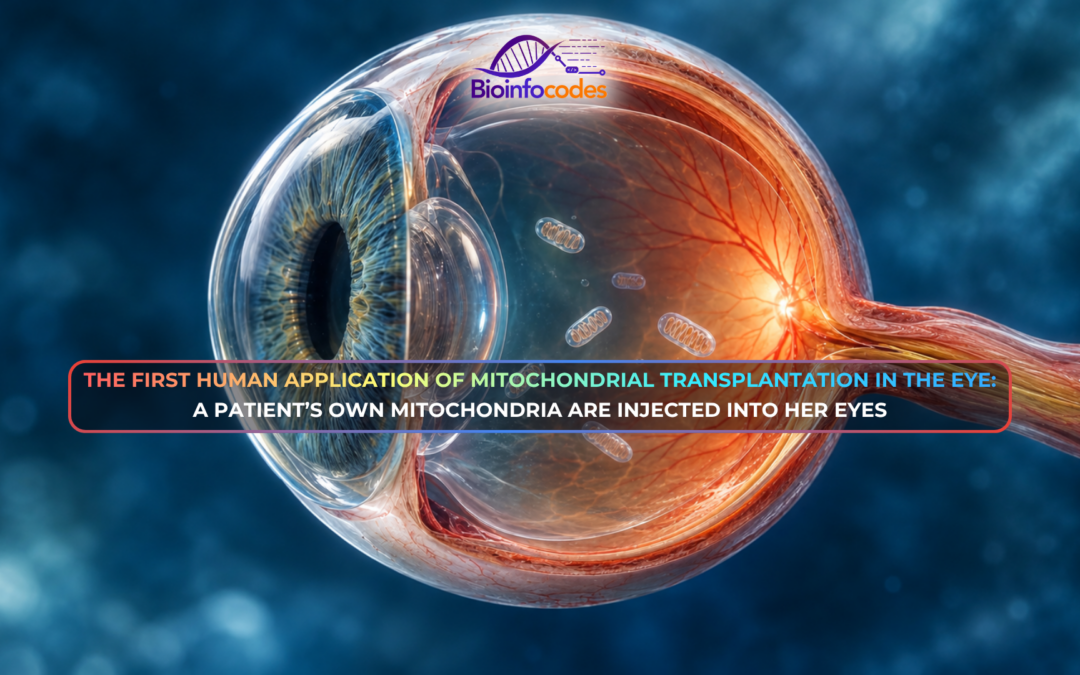
Mitochondrial transplantation is being explored as a potential approach for addressing tissue damage associated with mitochondrial dysfunction. Mitochondria (organelles that produce energy in cells) can be taken up by damaged cells and, in some cases, support cell survival and partially restore tissue function. Although previous human trials have evaluated mitochondrial transplantation in cardiac and cerebral tissues, the feasibility and safety of delivering isolated mitochondria into the eye remain an area of ongoing investigation.
The researchers aimed to assess the safety of injecting a patient’s own mitochondria into the eye while observing whether any functional responses could be detected. A brain bleed in February had injured a woman’s optic nerve and retina, causing nearly complete blindness and a total loss of pupil responses to light. Researchers aimed to assess the procedure’s safety, while investigating whether the intervention was followed by any changes in visual function. Retinal ganglion cells, which transmit visual signals from the retina to the brain through the optic nerve, were a key focus of the approach.
In May, the researchers received emergency authorization from the US Food and Drug Administration (FDA). They subsequently extracted mitochondria from the woman’s leg muscles and injected them into the vitreous fluid of her eyes. Safety was assessed by testing for signs of inflammation or tissue damage. Functional changes were tracked using pupillary light reflex assessments, low-vision evaluations, and electrical activity recordings in the visual cortex conducted before and after the transplantation.
Post-procedure assessments revealed no signs of intraocular inflammation or tissue damage in this patient, suggesting that the procedure was well tolerated. Following the intervention, the patient’s pupils showed a transient response to light, which subsided after approximately four weeks. Low-vision assessment noted shape and shadow perception in the left eye, and electrical activity was recorded in the visual cortex on days 3, 44, and 45 post-transplant (which was absent 27 days and 4 days prior to the procedure). However, the treatment did not restore the woman’s vision, and no direct evidence confirmed whether the injected mitochondria entered the cells or remained functionally active.
The findings, reported in a preprint that has not undergone peer review, provide preliminary evidence that intraocular mitochondrial injection was well tolerated in this patient, but do not establish therapeutic efficacy. Experts noted that observed pupillary and cortical changes could represent residual neuronal recovery or natural fluctuations rather than direct mitochondrial action. Researchers are also working with the FDA to develop a protocol for repeated mitochondrial injections.
Author: Elinsu Ak
Editor: Nur Tanem Altundaş
Reference: Naddaf, M. (2026). Patient’s own mitochondria injected into eyes in attempt to restore vision. Nature. https://doi.org/10.1038/d41586-026-02616-z
-Bioinfocodes Scientific News Service-
News articles prepared by our team members, reviewing and compiling scientific research
published in journals with and impact factor greater than 20 (click here for the list)