

Dilan AğırbaşA., Ebru KavaklıE.
RNA therapy is a type of treatment that regulates biological pathways with RNA molecules to remedy a particular disease1. Manipulating gene expression and producing therapeutic proteins are fundamental functions of RNA therapies. Moreover, it can convert the therapeutic proteins appropriate for cancers, infectious diseases, immune system disorders, and pathologies with defined genetic targets2. Compared to the drugs that can only target a limited number of proteins contributing to disease pathways, RNA-based therapeutics show promise in increasing the number of ‘druggable’ targets3. Inhibitors of mRNA translation (antisense), RNA interference (RNAi) agents, ribozymes, messenger RNA (mRNA), and aptamers are the main types of RNA therapeutics that will be clarified in this paper4,5.
RNAi (Small Interfering RNA, MicroRNA)
The internal cellular process that utilizes double-stranded RNAs (dsRNAs), which are a crucial trigger in the body’s natural immune system against viral infections, to silence genes in the gene expression is called RNAi6-8.
Small Interfering RNA (siRNA)
siRNAs that have 5′-phosphate/3′-hydroxyl ends and two 3′-overhang ribonucleotides on each duplex strand belong to the group of short dsRNAs9,10. Based on the complementarity of the sequences, siRNAs bind to their target transcript and bring out an RNAi response11. Dicer proteins associated with RNase III initiate the RNAi mechanism by cleaving a dsRNA into shorter duplexes12. RNA-induced silencing complex (RISC), a multi-protein component complex, binds to the resulting siRNAs13. The siRNA is then cleaved into strands within the RISC complex, and the stable 5′ end strand amalgamates with the active RISC complex14. The RISC complex located on the target mRNA is positioned by antisense single-stranded siRNA. Next, mRNA cleavage occurs by the action of the catalytic RISC protein, a member of the argonaute family (Ago2) (Figure 1)15. siRNAs have been used to study the effect of different oncogenic long non-coding RNAs (lncRNAs) on loss of function. As a result of the studies, it was found that siRNAs have a silencing property against lncRNA HOX transcript antisense RNA (HOTAIR), whose role is to foster metastasis in cell lung cancer 16.
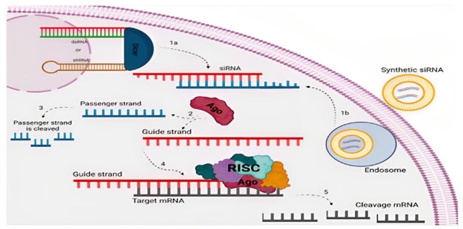
Figure 1. Mechanism of gene silencing via siRNA in eukaryotic cells with different pathways15. 1a) DICER cleaves dsRNA/shRNA into mature siRNA (Endogenous pathway). 1b) Synthetic siRNA enters via endosome, released into the cytoplasm (Exogenous pathway). 2) AGO protein separates strands. 3) The passenger strand is cleaved. 4) RISC complex selects guide strand for mRNA alignment. 5) mRNA is cleaved to silence the target gene.
miRNA
This RNA strain, which belongs to the class of non-coding RNA and is ~22-nt in length, controls gene expression and causes gene silencing by binding to target mRNAs after transcription17,18. Primary miRNA (pri-miRNA) is formed from miRNA genes transcribed by the enzyme RNA polymerase II in the nucleus. Then, RNase III Drosha and microprocessor complex subunit DiGeorge syndrome critical region 8 (DGCR8) cleave the pri-miRNA, resulting in a precursor miRNA (pre-miRNA) containing 20–25 nucleotides19,20. Subsequently, Exportin 5 delivers the pre-miRNA from the nucleus to the cytoplasm, and the Dicer enzyme converts the pre-miRNAs transported into the cytoplasm into an 18-25 nucleotide miRNA duplex21,22. The miRNA duplex then joins the RISC to create the miRISC complex. In contrast to the processing of siRNA, where the RISC’s AGO2 leads to the cleavage of the siRNA’s passenger strand, the miRNA duplex opens, releasing and discarding the passenger strand. miRISC complex is directed to target mRNAs by mature single-stranded miRNAs23. The miRNA connects to the target mRNAs, silencing the target gene with translational repression, degradation, and/or cleavage through partial complementary base pairing (Figure 2)24.
In the process of the growth and spread of tumors, miRNAs either behave as tumor suppressors or oncogenes. The research found that controlling miRNA modifications via miRNA mimics can restore the gene regulatory network and alter the phenotypes in cancer cells25. Numerous miRNA-targeted therapeutics are now in the clinical development stage. These include miR-122 antagonists that have entered phase II trials for treating hepatitis and a mimic of the tumor suppressor miRNA miR-34 that has reached phase I clinical trials26.
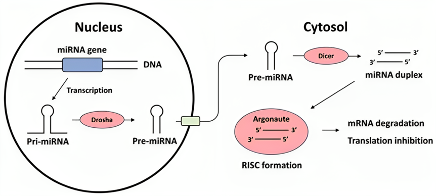
Figure 2. Biogenesis and mechanism of action of microRNAs24.
Antisense Oligonucleotides (ASOs)
ASOs are complementary nucleic acid fragments created to hybridize to a complementary endogenous pre-mRNA or mRNA through Watson-Crick base pairing27. There are two discrete ways that ASOs function. The first involves RNA degradation, while the second entails RNA inhibition or modulation via steric hindrance28-30.
In the RNA degradation, target RNA is bound by ASO, resulting in RNA/ASO duplexes. RNaseH1, an endonuclease that cleaves the phosphodiester bonds of RNA, is recruited by these duplexes. This process occurs for mRNA in the cytoplasm31,32. 5′-3′ exoribonucleases (XRNs) and the exosome complex cleave the RNA fragments after RNaseH1 cleavage. Researchers discovered that ASO gene silencing was abolished by observing that the pre-RNA levels of the same gene increased with increased transcription as the target mRNA in the ribosomes was cleaved by the recruited RNase H133. In the RNA steric hindrance, RNA/ASO duplexes are rendered resistant to endonuclease cleavage by added sugar modifications to phosphorothioate-modified ASOs34. Failure to assemble the initiation complex at the AUG start codon occurs because of the binding of such duplexes within the 5′ untranslated region of the mRNA, and mRNA translation is sterically stopped (Figure 3)35. Furthermore, by averting 5′ capping or directing splice site selection, steric hindrance can control how pre-mRNA is processed in the nucleus. Steric inhibitory ASOs can also be used to increase the expression of therapeutically effective proteins36. The interaction of the preinitiation complex with the primary open reading frame (ORF) is enhanced by ASOs that block upstream open reading frames in mRNA. In consequence, protein expression is increased35.
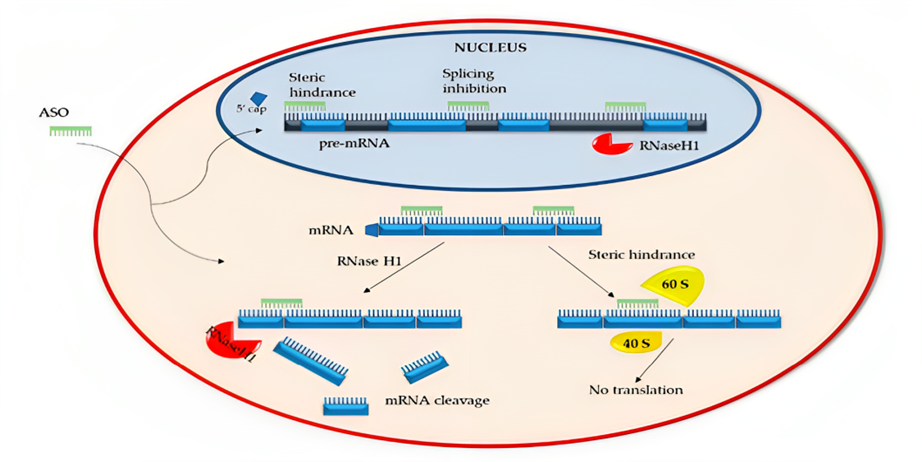
Figure 3. Mechanism of action of ASOs35. ASOs facilitate cleavage of pre/mRNAs via RNaseH1. Additionally, exon splicing, interaction of mRNA with ribosomal subunits (40S and 60S) and 5′ capping of the pre-RNA processes are blocked by ASOs.
Aptamers
Oligonucleotide sequences that behave similarly to monoclonal antibodies and are 25-80 bases in length are called Aptamers37. Usually, they bind to specific protein targets by transforming into various 3D structures38,39. A conceptually simple iterative selection procedure known as SELEX is used to isolate Aptamers from a pool of nucleic acids40-42.
The US Food and Drug Administration (FDA) has approved only one RNA-based aptamer drug to date. This drug is pegaptanib, a 28-nucleotide construct with two polyethylene glycol (PEG) fragments attached to the end43,44. To suppress cell proliferation, the main downstream effect of vascular endothelial growth factor (VEGF) signaling, pegaptanib binds to the 165 isoform of VEGF and blocks its interaction with the receptor protein (Figure 4)45. In the context of this effect, pegaptanib was designed as a therapeutic agent to treat wet-type (neovascular) age-related macular degeneration (AMD), an illness that causes damage to the central retina46,47. Pegaptanib is a good example of how RNA-based aptamers can be used as therapeutics, despite being infrequently used nowadays owing to competition from numerous antibody-based drugs with comparable efficacy48.
Ribozymes
Catalytic RNA molecules that can reduce the expression of targeted genes by breaking down certain RNA sequences are called ribozymes49. Although there are several classes of ribozymes, hammerhead ribozyme (HHRz) has received significant attention due to being one of the smallest ribozymes50.
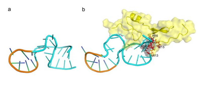
Figure 4. Interaction of Macugen (pegaptanib) with its target protein and its structural model45. a) Based on the secondary structure determined by NMR, the pegaptanib structure model was presented. b) Interface of target protein VEGF165 and aptamer. Yellow parts were used to depict the VEGF165 surface and structure. The blue areas are residues that have undergone conformational change on pegaptanib. The VEGF165 residues that were involved in the complex’s interaction were highlighted in red and identified in black (R13, R14, K15, H16).
The catalytic core (22 nt-long) that controls the cleavage reaction and three hybridizing helices, two of which surround the catalytic core and utilize Watson-Crick base pairing to bind the target RNA in an antisense approach are the parts that make up the HHRz51,52. Target RNA that contains NUH sequences, where N is any base and H is A, U, or C, is typically recognized by HHRz (Figure 5)53. Target sites with the GUA, GUC, GUU, CUC, and UUC sequences are preferred the most. By immediate 3′ cleavage of the selected RNA into the NUH sequence, hammerhead ribozymes’ primary mechanism of action in repressing target gene expression is activated. This action results in two shorter types of RNA that lack stabilizing sequences, undergo rapid degradation, and cause a progressive decrease in protein synthesis50,54. Compared to other antisense molecules in RNAi systems, a single ribozyme can recognize and bind more than one target mRNA55,56. Ribozymes typically mediate multiple-turnover and sequence-specific reactions, and both features make these molecules appropriate candidates for therapeutic use57. Targeting the 5′ URT of the hepatitis C virus (HCV) RNA genome, Heptazyme is one of the antiviral ribozymes that have made it to the clinical stage58. Despite showing success in clinical trials, additional research is needed to enhance the stability, effectiveness, and safety of ribozymes59.
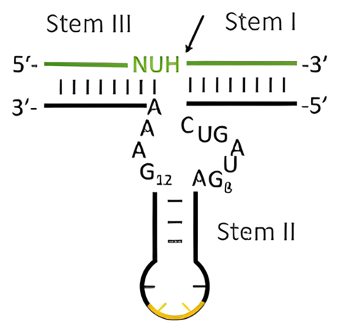
Figure 5. The secondary structure of a hammerhead ribozyme model53.
mRNA
It is a type of RNA obtained by copying from genomic DNA and whose function is to code proteins. The 5′ cap, 5′ untranslated region (UTR), coding region, 3′ UTR, and poly(A) tail are the components of mRNAs60,61.
Direct injection of mRNA into mice resulted in successful transfection and the production of an immune response, allowing the expression of therapeutic proteins61,62. This success led to the development of mRNA-based therapeutics. Infectious disease vaccines, genetic engineering, cancer immunotherapy, protein replacement therapy, and mRNA-based gene editing are the main application areas where mRNA is used today63. mRNA-based treatments can be divided into two categories according to their purposes: enzyme replacement therapy and vaccine against infectious diseases or cancer antigens60,64. Lipid nanoparticles (LNPs), which are lipid-based carrier systems, encapsulate mRNAs that will encode the target protein65. The resulting LNP-mRNAs reach the cytosol via endocytosis. Based on the mRNAs, proteins are translated inside the ribosomes (Figure 6)66. In vitro transcribed (IVT) mRNA protein replacement is functional in non-genetic disease conditions. IVT mRNA that encodes vascular endothelial growth factor-A (VEGF-A) could enhance blood flow, offering ischemic cardiovascular disease patients a regenerative treatment option67. For cancer immunotherapy, mRNA-based dendritic cell (DC) vaccines are applied. These vaccines can direct cytotoxic T lymphocytes and natural killer cells into powerful anti-tumor weapons that can target tumor cells68,69. In infectious diseases, IVT mRNA is delivered to the human body to translate antigenic proteins and stimulate the immune system against the pathogen70,71.
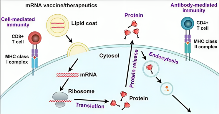
Figure 6. Peptide epitopes are cleaved by proteasome antigenic proteins66. To achieve cell-mediated immunity, CD8+ T cells are activated by loading these epitopes onto MHC I molecules. Secreted and endocytosed antigenic proteins are loaded onto MHC II located within endosomes. To help B cells produce antigen-specific antibodies for antibody-mediated immunity, MHC II-peptide complexes stimulate CD4+ T cells.
Several mRNA vaccine candidates for virus infections, including RSV (Respiratory Syncytial virus), influenza, Zika virus, and Cytomegalovirus (CMV), have entered clinical trials66.
Future Prospects and Conclusion
The field of RNA therapeutics is rapidly expanding and is still in its early stages72. RNA drugs are currently approved for cardiovascular, metabolic, liver, infectious, neurological, neuromuscular, renal, and eye diseases, and therapeutic intervention has improved73,74. The number of RNA medications in development and clinical trials is increasing quickly75. As a result of clinical studies, it was discovered that ASOs demonstrate a high affinity toward targets based on one-dimensional sequence matches76. Moreover, numerous ASO medications are currently being tested for genetic diseases and preventing complications from disease77. The ASO drugs mipomersen, eteplirsen nusinersen, inotersen, golodirsen, viltolarsen, and casimersen, and the siRNA drugs patisiran, givosiran and
lumasiran are among the therapeutics that have been successfully marketed since 201378. Researchers expect that siRNA and mRNA will soon be utilized in clinical trials to treat respiratory diseases owing to recent successes with siRNA therapeutics and technological advancements in RNA modification79. RNA therapeutics are poised to make an industry impact in a way diseases can be treated, thanks to approved treatment modalities in each of the major RNA therapeutic categories and significant research to improve their clinical use80.
Drugs and treatment methods mentioned in the review article are based only on the information obtained from the articles. Please consult a specialist physician for diagnosis, treatment, and drug use.
References
- Kim, Y.-K. (2022). RNA therapy: rich history, various applications and unlimited future prospects. Experimental & Molecular Medicine, 54(4), 455–465. https://doi.org/10.1038/s12276-022-00757-5
- Paunovska, K., Loughrey, D., & Dahlman, J. E. (2022). Drug delivery systems for RNA therapeutics. Nature Reviews Genetics, 23(5), 265–280. https://doi.org/10.1038/s41576-021-00439-4
- Ning, B., Yu, D., & Yu, A.-M. (2019). Advances and challenges in studying noncoding RNA regulation of drug metabolism and development of RNA therapeutics. Biochemical Pharmacology, 169, 113638. https://doi.org/10.1016/j.bcp.2019.113638
- Ning, L., Liu, M., Gou, Y., Yang, Y., He, B., & Huang, J. (2022). Development and application of ribonucleic acid therapy strategies against COVID-19. International Journal of Biological Sciences, 18(13), 5070–5085. https://doi.org/10.7150/ijbs.72706
- Dammes, N., & Peer, D. (2020). Paving the Road for RNA Therapeutics. Trends in Pharmacological Sciences, 41(10), 755–775. https://doi.org/10.1016/j.tips.2020.08.004
- Zhu, Y., Zhu, L., Wang, X., & Jin, H. (2022). RNA-based therapeutics: an overview and prospectus. Cell Death & Disease, 13(7), 644. https://doi.org/10.1038/s41419-022-05075-2
- Liu, S., Jaouannet, M., Dempsey, D. A., Imani, J., Coustau, C., & Kogel, K.-H. (2020). RNA-based technologies for insect control in plant production. Biotechnology Advances, 39, 107463. https://doi.org/10.1016/j.biotechadv.2019.107463
- Xu, W., Jiang, X., & Huang, L. (2019). RNA Interference Technology. In Comprehensive Biotechnology (pp. 560–575). Elsevier. https://doi.org/10.1016/B978-0-444-64046-8.00282-2
- Garbo, S., Maione, R., Tripodi, M., & Battistelli, C. (2022). Next RNA Therapeutics: The Mine of Non-Coding. International Journal of Molecular Sciences, 23(13), 7471. https://doi.org/10.3390/ijms23137471
- Charbe, N. B., Amnerkar, N. D., Ramesh, B., Tambuwala, M. M., Bakshi, H. A., Aljabali, A. A. A., Khadse, S. C., Satheeshkumar, R., Satija, S., Metha, M., Chellappan, D. K., Shrivastava, G., Gupta, G., Negi, P., Dua, K., & Zacconi, F. C. (2020). Small interfering RNA for cancer treatment: overcoming hurdles in delivery. Acta Pharmaceutica Sinica B, 10(11), 2075–2109. https://doi.org/10.1016/j.apsb.2020.10.005
- Neumeier, J., & Meister, G. (2021). siRNA Specificity: RNAi Mechanisms and Strategies to Reduce Off-Target Effects. Frontiers in Plant Science, 11. https://doi.org/10.3389/fpls.2020.526455
- Almeida, M. V., Andrade-Navarro, M. A., & Ketting, R. F. (2019). Function and Evolution of Nematode RNAi Pathways. Non-Coding RNA, 5(1), 8. https://doi.org/10.3390/ncrna5010008
- Setten, R. L., Rossi, J. J., & Han, S. (2019). The current state and future directions of RNAi-based therapeutics. Nature Reviews Drug Discovery, 18(6), 421–446. https://doi.org/10.1038/s41573-019-0017-4
- Mahmoodi Chalbatani, G., Dana, H., Gharagouzloo, E., Grijalvo, S., Eritja, R., Logsdon, C. D., Memari, F., Miri, S. R., Rezvani Rad, M., & Marmari, V. (2019). <p>Small interfering RNAs (siRNAs) in cancer therapy: a nano-based approach</p>. International Journal of Nanomedicine, Volume 14, 3111–3128. https://doi.org/10.2147/IJN.S200253
- de Brito e Cunha, D., Frederico, A., Azamor, T., Melgaço, J., da Costa Neves, P., Bom, A., Tilli, T., & Missailidis, S. (2022). Biotechnological Evolution of siRNA Molecules: From Bench Tool to the Refined Drug. Pharmaceuticals, 15(5), 575. https://doi.org/10.3390/ph15050575
- Li, H., Cui, Z., Lv, X., Li, J., Gao, M., Yang, Z., Bi, Y., Zhang, Z., Wang, S., Li, S., Zhou, B., & Yin, Z. (2020). Long Non-coding RNA HOTAIR Function as a Competing Endogenous RNA for miR-149-5p to Promote the Cell Growth, Migration, and Invasion in Non-small Cell Lung Cancer. Frontiers in Oncology, 10. https://doi.org/10.3389/fonc.2020.528520
- Zhang, S., Cheng, Z., Wang, Y., & Han, T. (2021). The Risks of miRNA Therapeutics: In a Drug Target Perspective. Drug Design, Development and Therapy, Volume 15, 721–733. https://doi.org/10.2147/DDDT.S288859
- Menon, A., Abd-Aziz, N., Khalid, K., Poh, C. L., & Naidu, R. (2022). miRNA: A Promising Therapeutic Target in Cancer. International Journal of Molecular Sciences, 23(19), 11502. https://doi.org/10.3390/ijms231911502
- Gjorgjieva, M., Sobolewski, C., Dolicka, D., Correia de Sousa, M., & Foti, M. (2019). miRNAs and NAFLD: from pathophysiology to therapy. Gut, 68(11), 2065–2079. https://doi.org/10.1136/gutjnl-2018-318146
- de Rooij, L. A., Mastebroek, D. J., ten Voorde, N., van der Wall, E., van Diest, P. J., & Moelans, C. B. (2022). The microRNA Lifecycle in Health and Cancer. Cancers, 14(23), 5748. https://doi.org/10.3390/cancers14235748
- He, B., Zhao, Z., Cai, Q., Zhang, Y., Zhang, P., Shi, S., Xie, H., Peng, X., Yin, W., Tao, Y., & Wang, X. (2020). miRNA-based biomarkers, therapies, and resistance in Cancer. International Journal of Biological Sciences, 16(14), 2628–2647. https://doi.org/10.7150/ijbs.47203
- O’Brien, J., Hayder, H., Zayed, Y., & Peng, C. (2018). Overview of MicroRNA Biogenesis, Mechanisms of Actions, and Circulation. Frontiers in Endocrinology, 9. https://doi.org/10.3389/fendo.2018.00402
- Si, W., Shen, J., Zheng, H., & Fan, W. (2019). The role and mechanisms of action of microRNAs in cancer drug resistance. Clinical Epigenetics, 11(1), 25. https://doi.org/10.1186/s13148-018-0587-8
- Jung, H., Kim, J. S., Lee, K. H., Tizaoui, K., Terrazzino, S., Cargnin, S., Smith, L., Koyanagi, A., Jacob, L., Li, H., Hong, S. H., Yon, D. K., Lee, S. W., Kim, M. S., Wasuwanich, P., Karnsakul, W., Shin, J. Il, & Kronbichler, A. (2021). Roles of microRNAs in inflammatory bowel disease. International Journal of Biological Sciences, 17(8), 2112–2123. https://doi.org/10.7150/ijbs.59904
- Maximov, V. V., Akkawi, R., Khawaled, S., Salah, Z., Jaber, L., Barhoum, A., Or, O., Galasso, M., Kurek, K. C., Yavin, E., & Aqeilan, R. I. (2019). MiR‐16‐1‐3p and miR‐16‐2‐3p possess strong tumor suppressive and antimetastatic properties in osteosarcoma. International Journal of Cancer, 145(11), 3052–3063. https://doi.org/10.1002/ijc.32368
- Chakraborty, C., Sharma, A. R., Sharma, G., & Lee, S. S. (2021). Therapeutic advances of miRNAs: A preclinical and clinical update. In Journal of Advanced Research (Vol. 28, pp. 127–138). Elsevier B.V. https://doi.org/10.1016/j.jare.2020.08.012
- Amanat, M., Nemeth, C. L., Fine, A. S., Leung, D. G., & Fatemi, A. (2022). Antisense Oligonucleotide Therapy for the Nervous System: From Bench to Bedside with Emphasis on Pediatric Neurology. Pharmaceutics, 14(11), 2389. https://doi.org/10.3390/pharmaceutics14112389
- Di Fusco, D., Dinallo, V., Marafini, I., Figliuzzi, M. M., Romano, B., & Monteleone, G. (2019). Antisense Oligonucleotide: Basic Concepts and Therapeutic Application in Inflammatory Bowel Disease. Frontiers in Pharmacology, 10. https://doi.org/10.3389/fphar.2019.00305
- Rinaldi, C., & Wood, M. J. A. (2018). Antisense oligonucleotides: the next frontier for treatment of neurological disorders. Nature Reviews Neurology, 14(1), 9–21. https://doi.org/10.1038/nrneurol.2017.148
- Shadid, M., Badawi, M., & Abulrob, A. (2021). Antisense oligonucleotides: absorption, distribution, metabolism, and excretion. Expert Opinion on Drug Metabolism & Toxicology, 17(11), 1281–1292. https://doi.org/10.1080/17425255.2021.1992382
- Crooke, S. T., Baker, B. F., Crooke, R. M., & Liang, X. (2021). Antisense technology: an overview and prospectus. Nature Reviews Drug Discovery, 20(6), 427–453. https://doi.org/10.1038/s41573-021-00162-z
- Bennett, C. F. (2019). Therapeutic Antisense Oligonucleotides Are Coming of Age. Annual Review of Medicine, 70(1), 307–321. https://doi.org/10.1146/annurev-med-041217-010829
- Liang, X., Nichols, J. G., Hsu, C.-W., Vickers, T. A., & Crooke, S. T. (2019). mRNA levels can be reduced by antisense oligonucleotides via no-go decay pathway. Nucleic Acids Research, 47(13), 6900–6916. https://doi.org/10.1093/nar/gkz500
- Amanat, M., Nemeth, C. L., Fine, A. S., Leung, D. G., & Fatemi, A. (2022). Antisense Oligonucleotide Therapy for the Nervous System: From Bench to Bedside with Emphasis on Pediatric Neurology. Pharmaceutics, 14(11), 2389. https://doi.org/10.3390/pharmaceutics14112389
- Gagliardi, M., & Ashizawa, A. T. (2021). The Challenges and Strategies of Antisense Oligonucleotide Drug Delivery. Biomedicines, 9(4), 433. https://doi.org/10.3390/biomedicines9040433
- Curreri, A., Sankholkar, D., Mitragotri, S., & Zhao, Z. (2023). <scp>RNA</scp> therapeutics in the clinic. Bioengineering & Translational Medicine, 8(1). https://doi.org/10.1002/btm2.10374
- Juliana Nordin, F., Wan Ming, L., Yee Mun Teo, M., & Lian Aun In, L. (2023). Aptamer Development for Cancer Diagnostic. In Rapid Antigen Testing. IntechOpen. https://doi.org/10.5772/intechopen.1001613
- Ni, S., Zhuo, Z., Pan, Y., Yu, Y., Li, F., Liu, J., Wang, L., Wu, X., Li, D., Wan, Y., Zhang, L., Yang, Z., Zhang, B.-T., Lu, A., & Zhang, G. (2021). Recent Progress in Aptamer Discoveries and Modifications for Therapeutic Applications. ACS Applied Materials & Interfaces, 13(8), 9500–9519. https://doi.org/10.1021/acsami.0c05750
- Yang, S., Li, H., Xu, L., Deng, Z., Han, W., Liu, Y., Jiang, W., & Zu, Y. (2018). Oligonucleotide Aptamer-Mediated Precision Therapy of Hematological Malignancies. Molecular Therapy – Nucleic Acids, 13, 164–175. https://doi.org/10.1016/j.omtn.2018.08.023
- Stephens, M. (2022). The emerging potential of Aptamers as therapeutic agents in infection and inflammation. Pharmacology & Therapeutics, 238, 108173. https://doi.org/10.1016/j.pharmthera.2022.108173
- Razlansari, M., Jafarinejad, S., rahdar, A., Shirvaliloo, M., Arshad, R., Fathi-Karkan, S., Mirinejad, S., Sargazi, S., Sheervalilou, R., Ajalli, N., & Pandey, S. (2023). Development and classification of RNA aptamers for therapeutic purposes: an updated review with emphasis on cancer. Molecular and Cellular Biochemistry, 478(7), 1573–1598. https://doi.org/10.1007/s11010-022-04614-x
- Komarova, N., & Kuznetsov, A. (2019). Inside the Black Box: What Makes SELEX Better? Molecules, 24(19), 3598. https://doi.org/10.3390/molecules24193598
- Egli, M., & Manoharan, M. (2023). Chemistry, structure and function of approved oligonucleotide therapeutics. Nucleic Acids Research, 51(6), 2529–2573. https://doi.org/10.1093/nar/gkad067
- Shahryari, A., Saghaeian Jazi, M., Mohammadi, S., Razavi Nikoo, H., Nazari, Z., Hosseini, E. S., Burtscher, I., Mowla, S. J., & Lickert, H. (2019). Development and Clinical Translation of Approved Gene Therapy Products for Genetic Disorders. Frontiers in Genetics, 10. https://doi.org/10.3389/fgene.2019.00868
- Zhang, N., Chen, Z., Liu, D., Jiang, H., Zhang, Z.-K., Lu, A., Zhang, B.-T., Yu, Y., & Zhang, G. (2021). Structural Biology for the Molecular Insight between Aptamers and Target Proteins. International Journal of Molecular Sciences, 22(8), 4093. https://doi.org/10.3390/ijms22084093
- Kaur, H., Bruno, J. G., Kumar, A., & Sharma, T. K. (2018). Aptamers in the Therapeutics and Diagnostics Pipelines. Theranostics, 8(15), 4016–4032. https://doi.org/10.7150/thno.25958
- Israilevich, R., Mahmoudzadeh, R., Salabati, M., & Xu, D. (2021). Narrative review-drug delivery in age-related macular degeneration. Annals of Eye Science, 6, 33–33. https://doi.org/10.21037/aes-21-8
- Niederlender, S., Fontaine, J.-J., & Karadjian, G. (2021). Potential applications of aptamers in veterinary science. Veterinary Research, 52(1), 79. https://doi.org/10.1186/s13567-021-00948-4
- Chi Wong, B., Shahid, U., & Siew Tan, H. (2023). Ribozymes as Therapeutic Agents against Infectious Diseases. In RNA Therapeutics – History, Design, Manufacturing, and Applications. IntechOpen. https://doi.org/10.5772/intechopen.107141
- Asha, K., Kumar, P., Sanicas, M., Meseko, C., Khanna, M., & Kumar, B. (2018). Advancements in Nucleic Acid Based Therapeutics against Respiratory Viral Infections. Journal of Clinical Medicine, 8(1), 6. https://doi.org/10.3390/jcm8010006
- Wyszko, E., Popenda, M., Gudanis, D., Sarzyńska, J., Belter, A., Perrigue, P., Skowronek, P., Rolle, K., & Barciszewski, J. (2021). The model structure of the hammerhead ribozyme formed by RNAs of reciprocal chirality. Bioscience Reports, 41(1). https://doi.org/10.1042/BSR20203424
- Zhao, Y., Shu, R., & Liu, J. (2021). The development and improvement of ribonucleic acid therapy strategies. Molecular Therapy – Nucleic Acids, 26, 997–1013. https://doi.org/10.1016/j.omtn.2021.09.002
- Czapik, T., Piasecka, J., Kierzek, R., & Kierzek, E. (2022). Structural variants and modifications of hammerhead ribozymes targeting influenza A virus conserved structural motifs. Molecular Therapy – Nucleic Acids, 29, 64–74. https://doi.org/10.1016/j.omtn.2022.05.035
- Panda, K., Alagarasu, K., & Parashar, D. (2021). Oligonucleotide-Based Approaches to Inhibit Dengue Virus Replication. Molecules, 26(4), 956. https://doi.org/10.3390/molecules26040956
- Dönmüş, B., Ünal, S., Kirmizitaş, F. C., & Türkoğlu Laçin, N. (2021). Virus-associated ribozymes and nano carriers against COVID-19. Artificial Cells, Nanomedicine, and Biotechnology, 49(1), 204–218. https://doi.org/10.1080/21691401.2021.1890103
- Pandey, M., Ojha, D., Bansal, S., Rode, A. B., & Chawla, G. (2021). From bench side to clinic: Potential and challenges of RNA vaccines and therapeutics in infectious diseases. Molecular Aspects of Medicine, 81, 101003. https://doi.org/10.1016/j.mam.2021.101003
- Reza, Md. S., Mim, F., Quader, M. R., Khan, M. J. R., Hossain, Md. S., Uddin, K. R., Akter, S., Rahman, S., Roy, S., & Rumman, Md. A. (2021). The Possibility of Nucleic Acids to Act as Anti-Viral Therapeutic Agents—A Review. Open Journal of Medical Microbiology, 11(03), 198–248. https://doi.org/10.4236/ojmm.2021.113015
- Zhou, L.-Y., Qin, Z., Zhu, Y.-H., He, Z.-Y., & Xu, T. (2019). Current RNA-based Therapeutics in Clinical Trials. Current Gene Therapy, 19(3), 172–196. https://doi.org/10.2174/1566523219666190719100526
- Volpini, L., Monaco, F., Santarelli, L., Neuzil, J., & Tomasetti, M. (2023). Advances in RNA cancer therapeutics: New insight into exosomes as miRNA delivery. Aspects of Molecular Medicine, 1, 100005. https://doi.org/10.1016/j.amolm.2023.100005
- Zogg, H., Singh, R., & Ro, S. (2022). Current Advances in RNA Therapeutics for Human Diseases. International Journal of Molecular Sciences, 23(5), 2736. https://doi.org/10.3390/ijms23052736
- Qin, S., Tang, X., Chen, Y., Chen, K., Fan, N., Xiao, W., Zheng, Q., Li, G., Teng, Y., Wu, M., & Song, X. (2022). mRNA-based therapeutics: powerful and versatile tools to combat diseases. Signal Transduction and Targeted Therapy, 7(1), 166. https://doi.org/10.1038/s41392-022-01007-w
- Sun, H., Zhang, Y., Wang, G., Yang, W., & Xu, Y. (2023). mRNA-Based Therapeutics in Cancer Treatment. Pharmaceutics, 15(2), 622. https://doi.org/10.3390/pharmaceutics15020622
- Xu, S., Yang, K., Li, R., & Zhang, L. (2020). mRNA Vaccine Era—Mechanisms, Drug Platform and Clinical Prospection. International Journal of Molecular Sciences, 21(18), 6582. https://doi.org/10.3390/ijms21186582
- Zhang, G., Tang, T., Chen, Y., Huang, X., & Liang, T. (2023). mRNA vaccines in disease prevention and treatment. Signal Transduction and Targeted Therapy, 8(1), 365. https://doi.org/10.1038/s41392-023-01579-1
- Trepotec, Z., Lichtenegger, E., Plank, C., Aneja, M. K., & Rudolph, C. (2019). Delivery of mRNA Therapeutics for the Treatment of Hepatic Diseases. Molecular Therapy, 27(4), 794–802. https://doi.org/10.1016/j.ymthe.2018.12.012
- Li, D., Liu, C., Li, Y., Tenchov, R., Sasso, J. M., Zhang, D., Li, D., Zou, L., Wang, X., & Zhou, Q. (2023). Messenger RNA-Based Therapeutics and Vaccines: What’s beyond COVID-19? ACS Pharmacology & Translational Science, 6(7), 943–969. https://doi.org/10.1021/acsptsci.3c00047
- Gan, L.-M., Lagerström-Fermér, M., Carlsson, L. G., Arfvidsson, C., Egnell, A.-C., Rudvik, A., Kjaer, M., Collén, A., Thompson, J. D., Joyal, J., Chialda, L., Koernicke, T., Fuhr, R., Chien, K. R., & Fritsche-Danielson, R. (2019). Intradermal delivery of modified mRNA encoding VEGF-A in patients with type 2 diabetes. Nature Communications, 10(1), 871. https://doi.org/10.1038/s41467-019-08852-4
- Gu, Y., Duan, J., Yang, N., Yang, Y., & Zhao, X. (2022). mRNA vaccines in the prevention and treatment of diseases. MedComm, 3(3). https://doi.org/10.1002/mco2.167
- Weng, Y., Li, C., Yang, T., Hu, B., Zhang, M., Guo, S., Xiao, H., Liang, X.-J., & Huang, Y. (2020). The challenge and prospect of mRNA therapeutics landscape. Biotechnology Advances, 40, 107534. https://doi.org/10.1016/j.biotechadv.2020.107534
- Wei, H.-H., Zheng, L., & Wang, Z. (2023). mRNA therapeutics: New vaccination and beyond. Fundamental Research. https://doi.org/10.1016/j.fmre.2023.02.022
- Qureischi, M., Mohr, J., Arellano-Viera, E., Knudsen, S. E., Vohidov, F., & Garitano-Trojaola, A. (2022). mRNA-based therapies: Preclinical and clinical applications (pp. 1–54). https://doi.org/10.1016/bs.ircmb.2022.04.007
- Zhang, C., & Zhang, B. (2023). RNA therapeutics: updates and future potential. Science China Life Sciences, 66(1), 12–30. https://doi.org/10.1007/s11427-022-2171-2
- Sasso, J. M., Ambrose, B. J. B., Tenchov, R., Datta, R. S., Basel, M. T., DeLong, R. K., & Zhou, Q. A. (2022). The Progress and Promise of RNA Medicine─An Arsenal of Targeted Treatments. Journal of Medicinal Chemistry, 65(10), 6975–7015. https://doi.org/10.1021/acs.jmedchem.2c00024
- Lundstrom, K. (2018). Latest development on RNA-based drugs and vaccines. Future Science OA, 4(5), FSO300. https://doi.org/10.4155/fsoa-2017-0151
- Damase, T. R., Sukhovershin, R., Boada, C., Taraballi, F., Pettigrew, R. I., & Cooke, J. P. (2021). The Limitless Future of RNA Therapeutics. Frontiers in Bioengineering and Biotechnology, 9. https://doi.org/10.3389/fbioe.2021.628137
- Xiong, H., Veedu, R. N., & Diermeier, S. D. (2021). Recent Advances in Oligonucleotide Therapeutics in Oncology. International Journal of Molecular Sciences, 22(7), 3295. https://doi.org/10.3390/ijms22073295
- Thakur, S., Sinhari, A., Jain, P., & Jadhav, H. R. (2022). A perspective on oligonucleotide therapy: Approaches to patient customization. Frontiers in Pharmacology, 13. https://doi.org/10.3389/fphar.2022.1006304
- Yu, A.-M., & Tu, M.-J. (2022). Deliver the promise: RNAs as a new class of molecular entities for therapy and vaccination. Pharmacology & Therapeutics, 230, 107967. https://doi.org/10.1016/j.pharmthera.2021.107967
- Chow, M. Y. T., Qiu, Y., & Lam, J. K. W. (2020). Inhaled RNA Therapy: From Promise to Reality. Trends in Pharmacological Sciences, 41(10), 715–729. https://doi.org/10.1016/j.tips.2020.08.002
- Curreri, A., Sankholkar, D., Mitragotri, S., & Zhao, Z. (2023). <scp>RNA</scp> therapeutics in the clinic. Bioengineering & Translational Medicine, 8(1). https://doi.org/10.1002/btm2.10374